PocketAnatomy® is a registered brand name owned by © eMedia Interactive Ltd, 2009-2022.
iPhone, iPad, iPad Pro and Mac are trademarks of Apple Inc., registered in the U.S. and other countries. App Store is a service mark of Apple Inc.
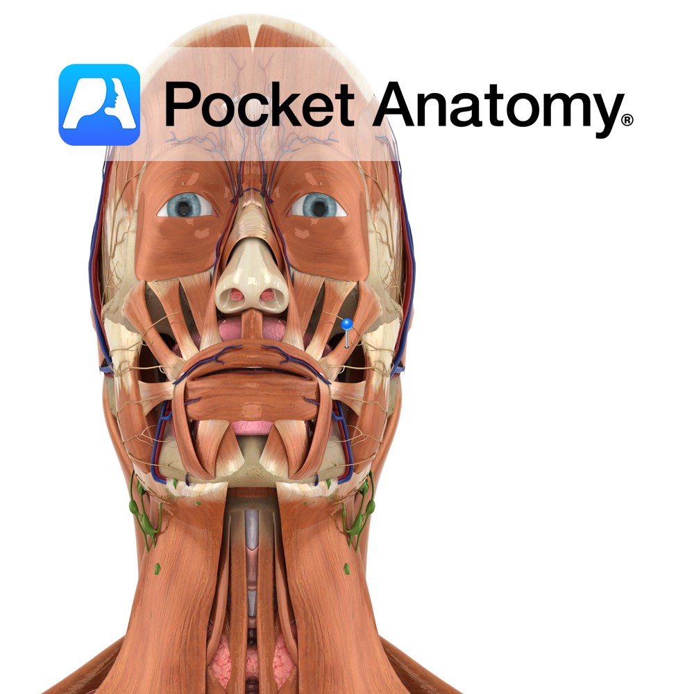
Anatomy Origin: Posterior part of the lateral surface of the zygomatic bone. Insertion: Angle of mouth where its fibres blend with those of levator anguli oris, depressor anguli oris and orbicularis oris. Key Relations: Arises posterior to orbicularis oculi and runs downwards and blends with orbicularis oris before inserting on the angle of the mouth.
- Published in Pocket Anatomy Pins
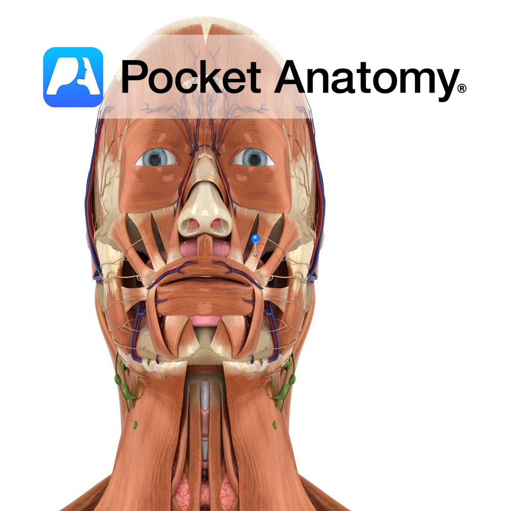
Anatomy Origin: Anterior part of the lateral surface of the zygomatic bone. Insertion: Upper lip, medial to the corner of the mouth. Key Relations: Arises anterior to the origin of zygomaticus major on the zygomatic bone. Functions Assists in deepening or elevating the nasolabial furrow e.g. when making expressions of disdain or contempt.. Supply Nerve
- Published in Pocket Anatomy Pins
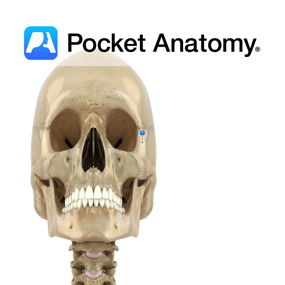
Anatomy One of four processes of zygomatic bone (malar, cheekbone). Is thick, extends up, articulates with zygomatic process of frontal bone and with it, forms strong lateral frame of orbit. Vignette Zygon (Greek): yoke or crossbar. Interested in taking our award-winning Pocket Anatomy app for a test drive?
- Published in Pocket Anatomy Pins

Anatomy Joins with the zygomatic process of the temporal bone, to form the zygomatic arch. Allows tendon of temporalis to pass medially (and lateral to temporal and sphenoid), down and forward to coronoid process of mandible. Vignette Tripod fracture; common facial injury which essentially amounts to separation of zygoma from rest of face; fractures of
- Published in Pocket Anatomy Pins
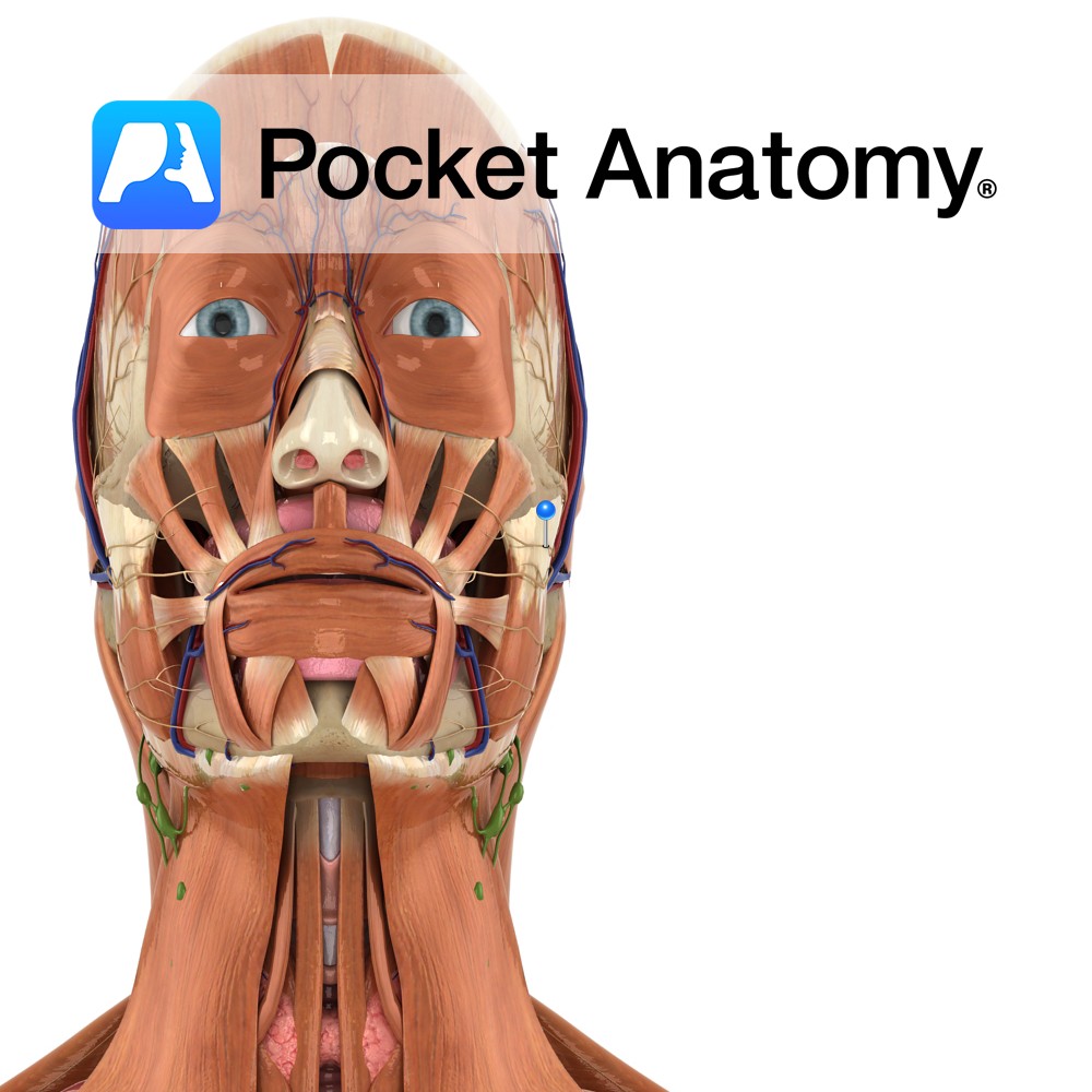
Anatomy Branch of the facial nerve (also known as the seventh cranial nerve). Facial nerve: Has a motor and sensory origin that join together to form the nerve. It passes through the internal auditory meatus through the facial canal and finally exits from the stylomastoid foramen, and into the parotid gland where it divides into
- Published in Pocket Anatomy Pins
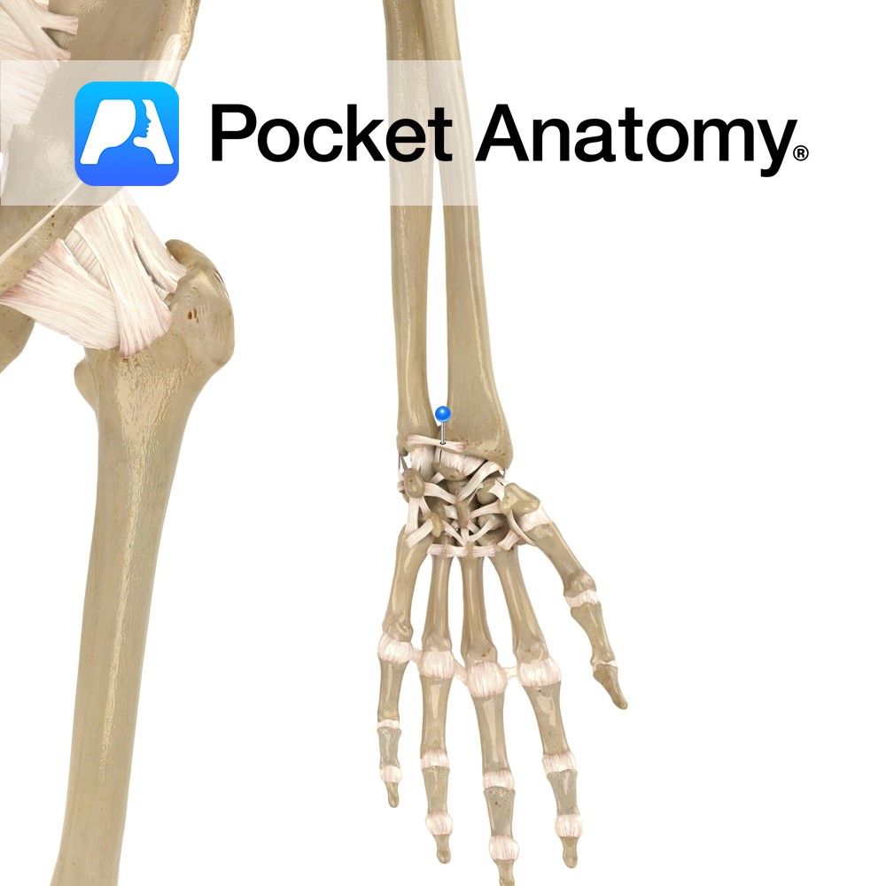
Anatomy Attaches from the ulna notch on the radius to the front of the distal part of the ulna. Functions Helps to hold the distal radioulnar joint together. Interested in taking our award-winning Pocket Anatomy app for a test drive?
- Published in Pocket Anatomy Pins
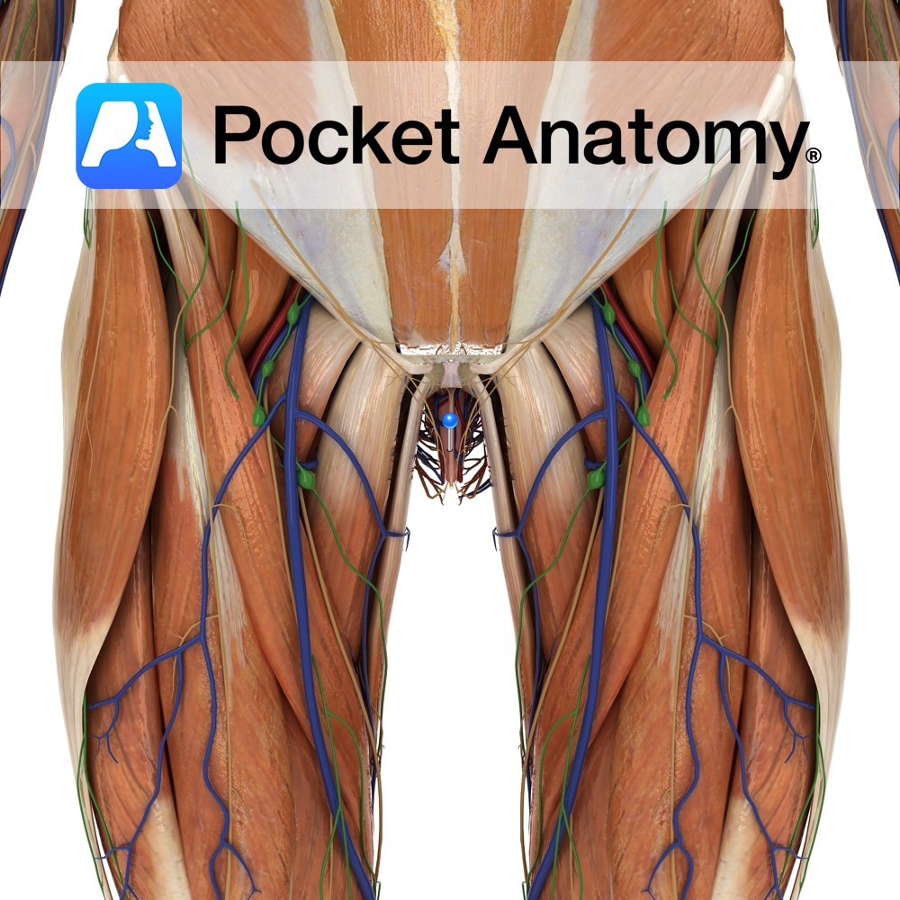
Anatomy External female genitalia (including mons pubis, prepuce, clitoris, labia majora/minora, urethral/vaginal orifices, hymen). Interested in taking our award-winning Pocket Anatomy app for a test drive?
- Published in Pocket Anatomy Pins
.jpg)
Motion The wrist (radiocarpal) joint is a multiaxial synovial ellipsoid joint. The distal end of the radius and the articular disc articulate with the proximal row of carpal bones (scaphoid, lunate and triquetral). The triangular articular disc is interposed between the distal end of the ulna and the carpus. The movements possible at the wrist
- Published in Pocket Anatomy Pins
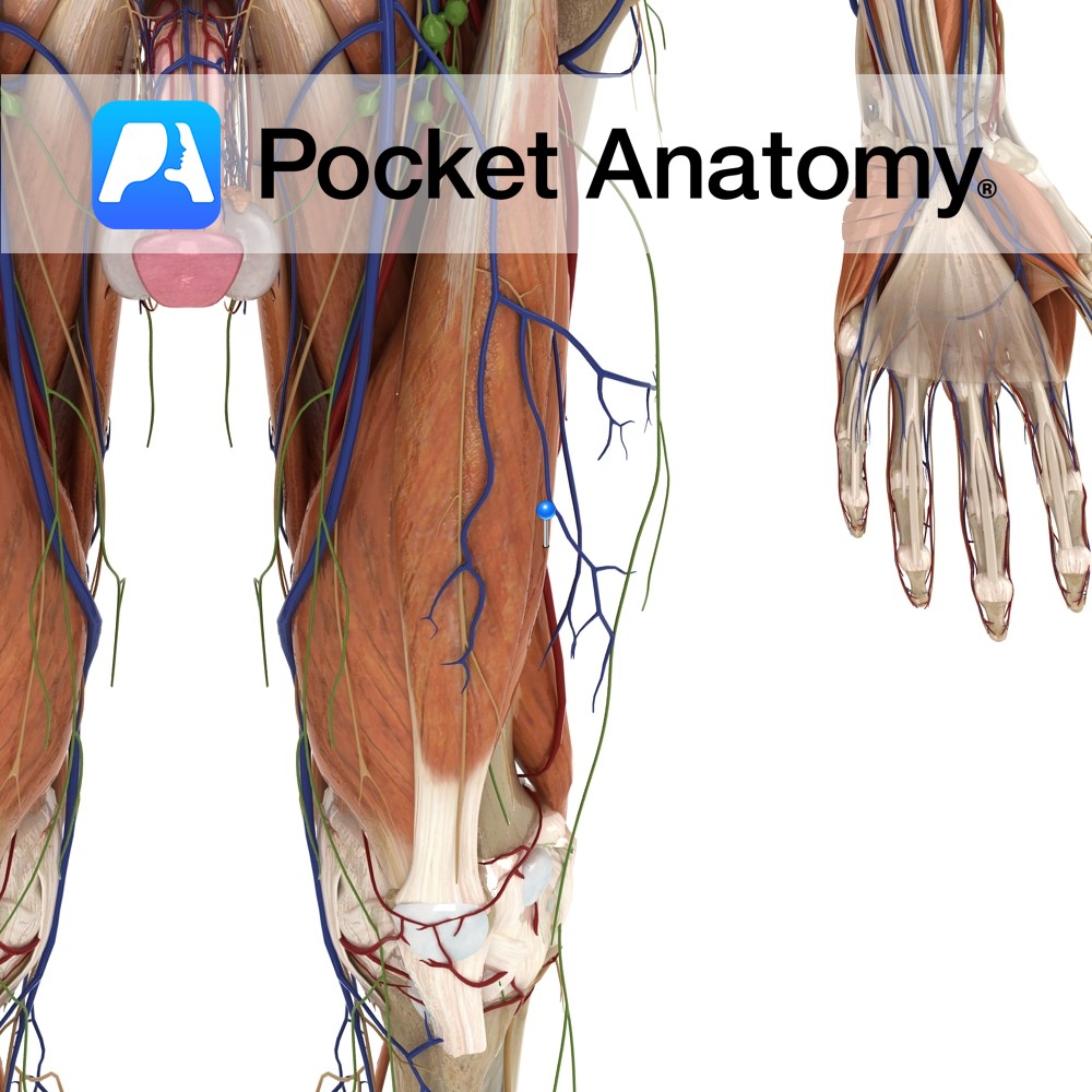
Anatomy Origin: Anterior and lateral surfaces of the proximal two-thirds of the femoral shaft. Insertion: Lateral margin and base of the patella via the quadriceps femoris tendon. The quadriceps femoris tendon is functionally continuous with the patellar ligament which runs from the apex of the patella to the tibial tuberosity. Key Relations: One of the
- Published in Pocket Anatomy Pins
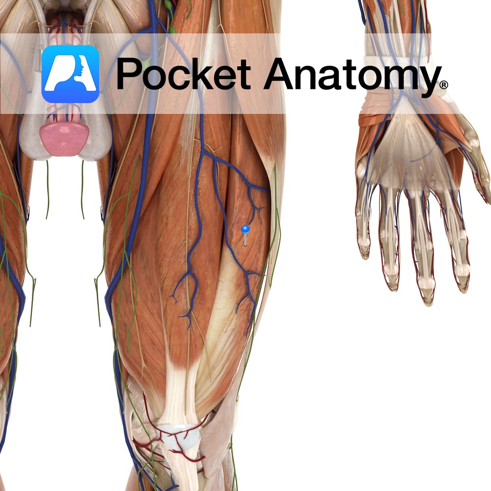
Anatomy Origin: Greater trochanter and lateral lip of linea aspera of femur. Insertion: Base of the patella via the quadriceps femoris tendon. The quadriceps femoris tendon is functionally continuous with the patellar ligament which runs from the apex of the patella to the tibial tuberosity. Key Relations: -One of the five muscles of the anterior
- Published in Pocket Anatomy Pins