Anatomy
Origin:
Long head: Superomedial area of ischial tuberosity (with semitendinosus).
Short head: Lateral lip of linea aspera and upper lateral supracondylar line of femur and lateral intermuscular septum.
Insertion:
The two heads insert by a common tendon into the lateral surface of head of fibula.
Key Relations:
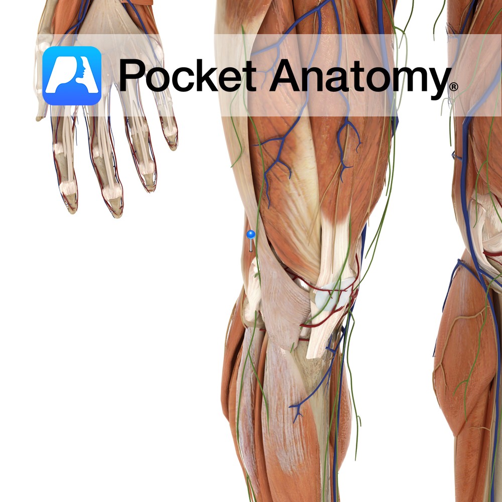
-One of the three muscles of the posterior compartment of the thigh.
-Lies anterolateral to semitendinosus and semimembranosus.
-Forms the superolateral boundary of the popliteal fossa.
Functions
-Flexes the leg at the knee joint.
-Extends the thigh at the hip joint (Long head only).
-Laterally rotates thigh at the hip joint when the hip is extended.
-Laterally rotates the leg at the knee joint when the knee is semi-flexed.
Supply
Nerve Supply:
Long head: Tibial division of sciatic nerve (L5, S1, S2)
Short head: Common peroneal (fibular) division of sciatic nerve (L5, S1, S2)
Blood Supply:
-Perforating arteries (branches of deep artery of thigh)
-Superior muscular branches of popliteal artery.
Clinical
‘Hamstring syndrome’ presents as pain near the ischial tuberosity thought to be caused by tendinitis at the origin of the hamstrings, in some cases compression of the sciatic nerve may also be involved. It is common amongst athletes, especially those partaking in sports that involve sprinting or short bursts of acceleration. The pain associated with ‘hamstring syndrome’ often radiates down the posterior thigh and is exacerbated by actions that stretch the hamstrings.
In addition the clinical examination of ‘hamstring syndrome’ may reveal tenderness over the ischial tuberosity and percussion in that region may cause the pain to radiate down the posterior thigh in a pattern determined by the sciatic nerve. ‘Hamstring syndrome’ can be treated with rest, NSAIDs and steroid injections. Surgical exploration of the sciatic nerve may be required to release various bands.
Interested in taking our award-winning Pocket Anatomy app for a test drive?
![]()