Anatomy
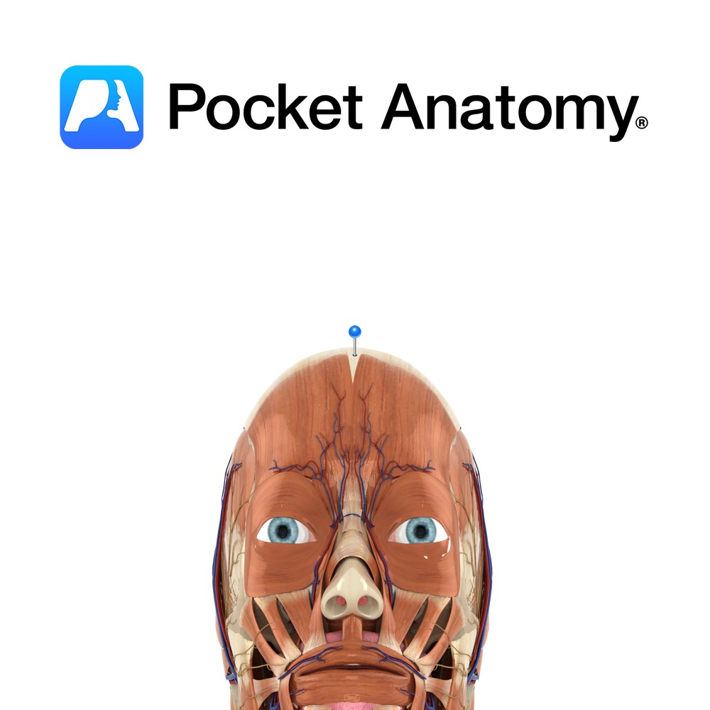
Layer of tough, dense, fibrous tissue that covers the upper portion of the cranium. Attaches posterior to occipitalis, anteriorly to frontalis, and laterally to auriculares anterior and superior. Posteriorly and laterally, its fibers are continued to the external occipital protuberance and zygomatic arch, respectively.
Dorsally, it is firmly connected to the skin by the superficial fascial layer; ventrally, it is loosely connected to the pericranium by cellular tissue.
Clinical
Anteriorly, the aponeurosis extends to the upper eyelids due to the lack of a bony insertion. This provides a potential subaponeurotic space where blood and fluids could migrate from the scalp to the upper eyelids.
Blood vessels are firmly attached to the superficial fascial layer, therefore cut ends of vessels do not easily withdraw. A small scalp laceration can cause large blood loss.
Interested in taking our award-winning Pocket Anatomy app for a test drive?
![]()