Anatomy
Course
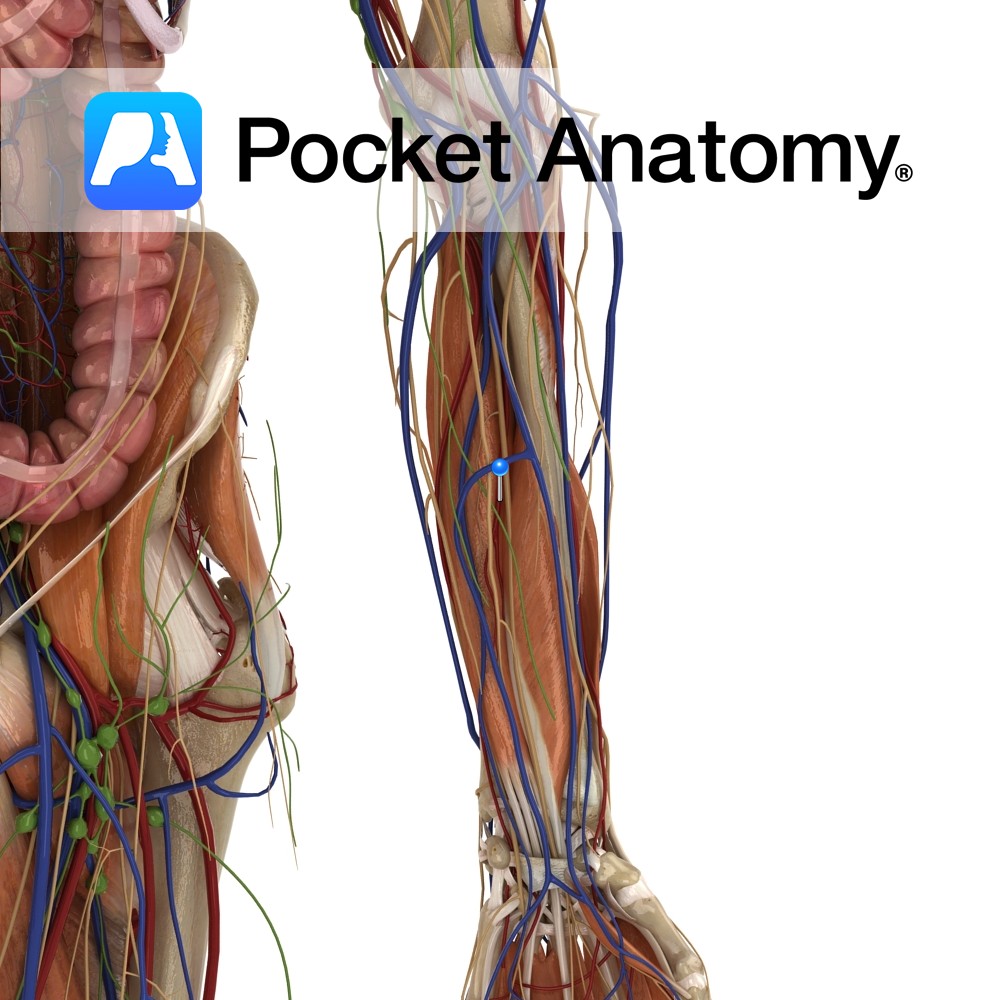
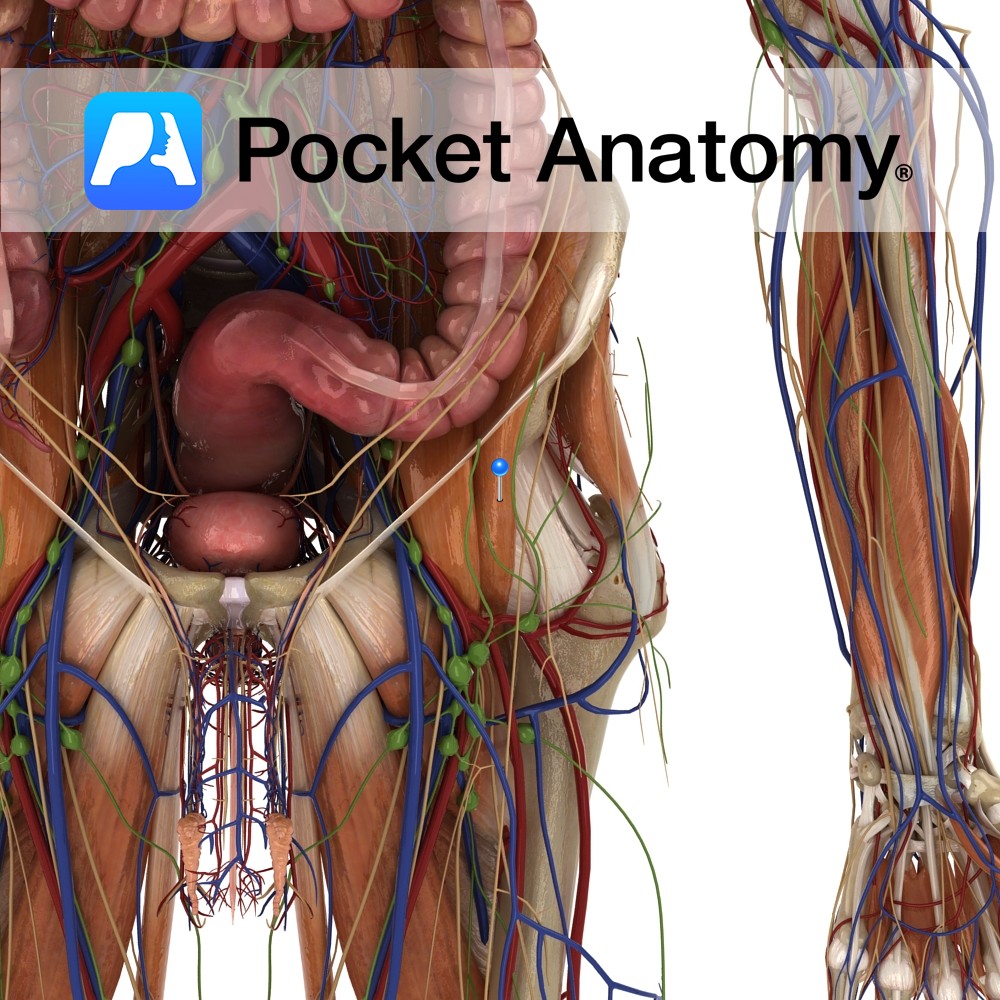
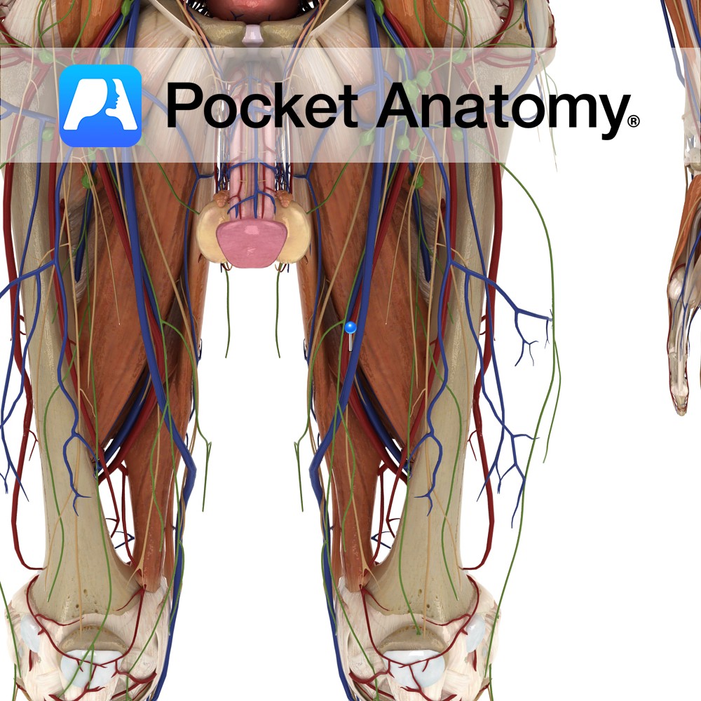
The external iliac artery becomes the femoral artery as it passes below the inguinal ligament and enters the femoral triangle, where it then descends in the adductor canal. It eventually exits via the adductor hiatus of the adductor magnus muscle, where it becomes the popliteal artery.
Supply
Supplies cutaneous regions of the lower abdomen, perineum and upper thigh as well as muscles in the thigh.
Clinical
The femoral artery is a common site for Peripheral Artery Disease (PAD). This is most commonly caused by a build up of atherosclerotic plaque in the femoral artery until blood flow to the lower leg is extremely impaired. At this stage a patient might experience cold feet, gangrene, loss of pulses in legs or feet as well as pain in the legs while lying down. One intervention is a femoral popliteal bypass where a blood vessel or vein graft can be used to connect the artery above and below the blockage.
Interested in taking our award-winning Pocket Anatomy app for a test drive?
![]()