-tertius.jpg)
Anatomy
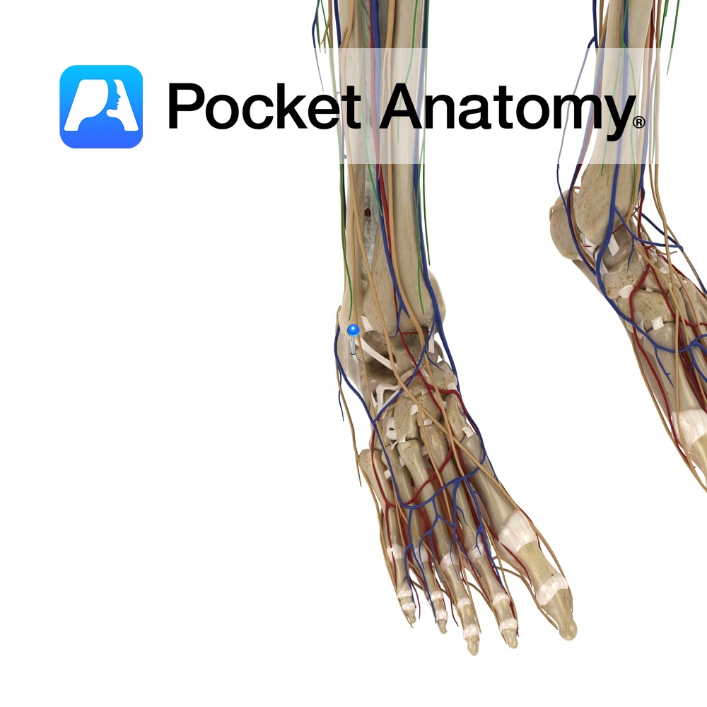
Origin:
Distal quarter of the anterior surface of the fibula and the interosseous membrane.
Insertion:
Dorsal surface of the base of the 5th metatarsal.
Key relations:
-One of the four muscles of the anterior compartment of the leg.
-The fibularis tertius tendon passes posterior to the extensor retinaculae. It crosses anterior to the ankle joint lateral to extensor digitorum longus.
Functions
-Everts the foot.
-Dorsiflexes the ankle joint.
Supply
Nerve Supply:
Deep fibular (peroneal) nerve (L5, S1).
Blood Supply:
Anterior tibial artery.
Clinical
The common peroneal nerve may be damaged easily at the neck of the fibula resulting in loss of function of the dorsiflexors of the ankle. The patient may present with ‘foot-drop’ as a result and may circumduct the hip when walking, to compensate for the ‘foot-drop’.
Interested in taking our award-winning Pocket Anatomy app for a test drive?
![]()