Anatomy
Origin:
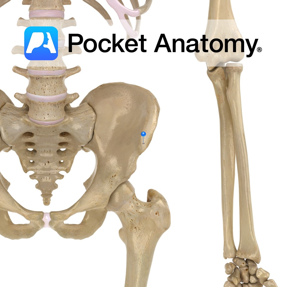
Humeral head: Medial epicondyle of humerus via the common flexor tendon.
Ulnar head: Medial margin of olecranon and upper two thirds of dorsal border of ulna by an aponeurosis.
Insertion:
Pisiform bone, then via pisohamate and pisometacarpal ligaments into the hook of the hamate and the fifth metacarpal.
Key Relations:
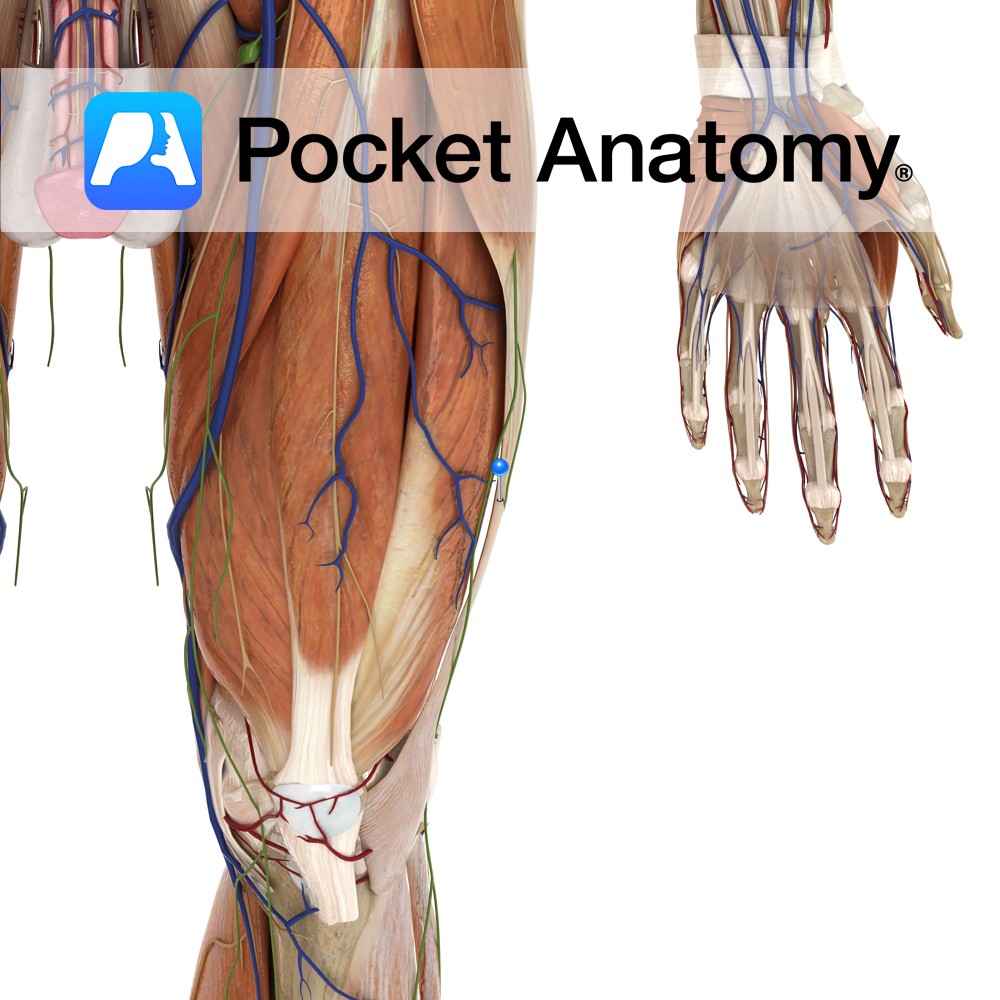
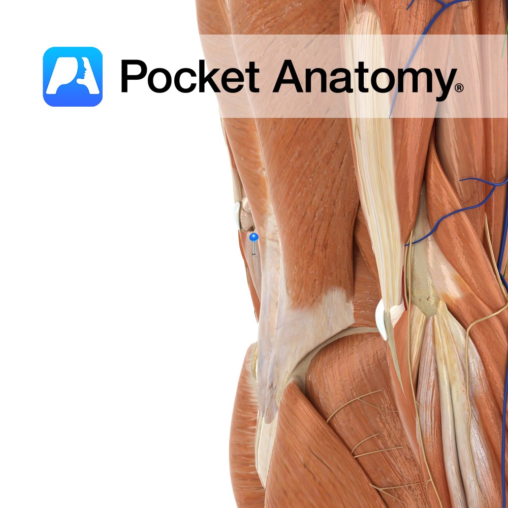
-The ulnar nerve courses between the two heads in the forearm before the two heads fuse just distal to the medial epicondyle of the humerus.
-One of the four muscles in the superficial anterior compartment of the forearm.
Functions
-Extends the hand at the wrist joint.
-Adducts the hand at the wrist joint (ulnar deviation) particularly when acting together with extensor carpi ulnaris.
e.g. as in pulling a rope towards you.
Supply
Nerve Supply:
Ulnar nerve (C7,C8).
Blood Supply:
Ulnar artery.
Clinical
Cubital tunnel syndrome is the second most common compression neuropathy behind carpal tunnel syndrome. One cause of cubital tunnel syndrome is the ulnar nerve becoming compressed between the two heads of flexor carpi ulnaris. Activities involving repeated flexion of the wrist, often combined with flexion of the fingers (flexor digitorum profundus forms the deep wall of the tunnel) can lead to the problem. One example of an activity involving these movements is when playing the guitar. For right handed people it would commonly affect the left arm, the side playing the fret board.
Interested in taking our award-winning Pocket Anatomy app for a test drive?
![]()
.jpg)