Anatomy
Origin:
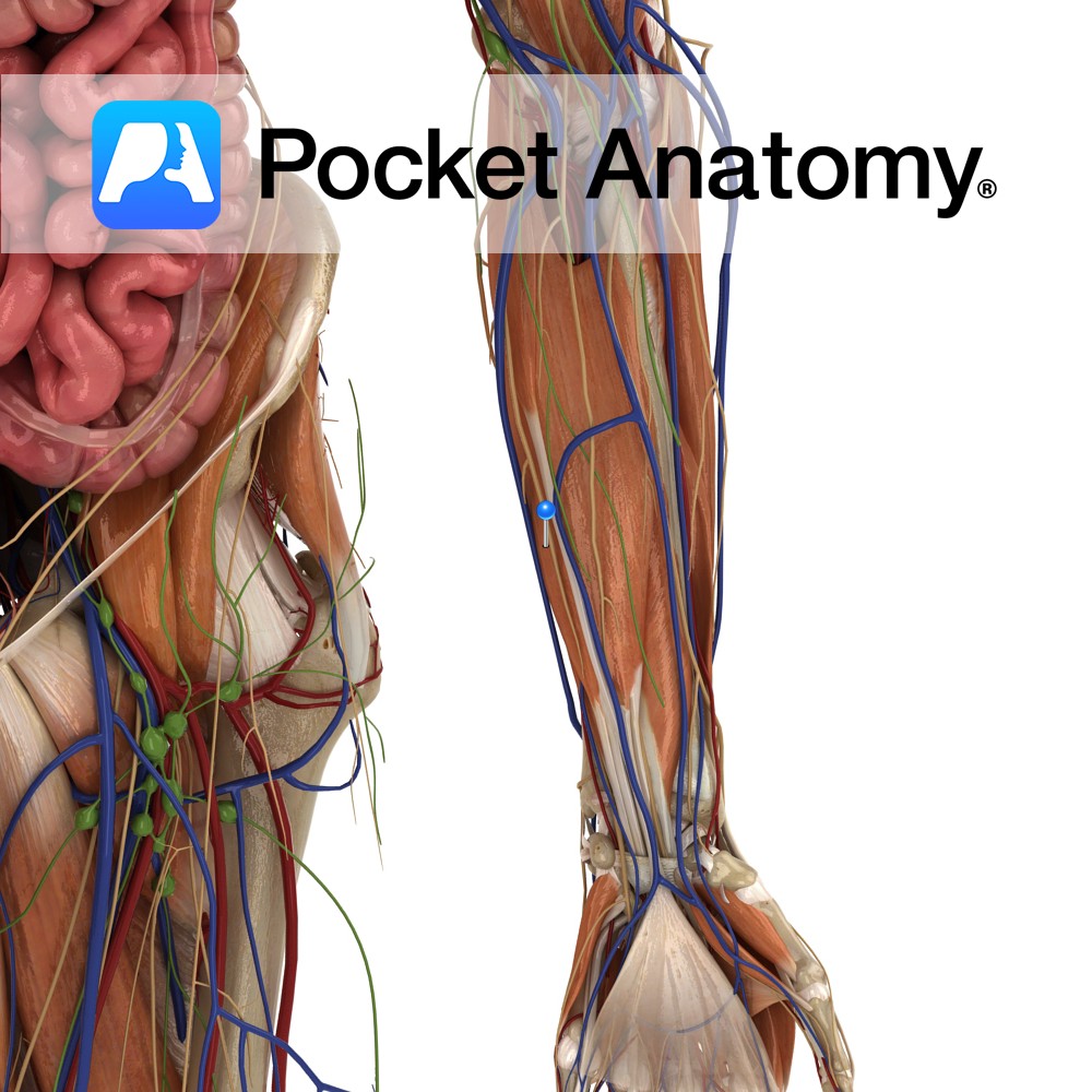
Humeroulnar head: Medial epicondyle of humerus via the common flexor tendon, ulnar collateral ligament and coronoid process of ulna.
Radial head: Superior half of anterior aspect of radius at anterior oblique line.
Insertion:
Four tendons of insertion attach to palmar surfaces of bodies of middle phalanges of the four fingers.
Key Relations:
-The tendons of insertion are split near their ends forming fibrous tunnels which allow the tendons of flexor digitorum profundus to reach the distal interphalangeal joints of the fingers. This split is known as camper’s chiasm.
-The four tendons of the muscle pass through the carpal tunnel as they enter the palm of the hand.
-The only muscle in the intermediate anterior compartment of the forearm.
Functions
-Flexor of the proximal interphalangeal joints of the fingers.
-Flexor of the metacarpophalangeal joints of the fingers
-Helps flex the hand at wrist joint
e.g. as in making a fist.
Supply
Nerve Supply:
Median nerve (C7,C8,T1).
Blood Supply:
Ulnar artery.
Clinical
Clinical testing is by flexing each finger at the proximal interphalangeal joint against resistance, while the remaining three fingers are held fully extended (this inactivates flexor digitorum profundus).
The tendons to the middle and ring fingers are more superficial than those to the little and index fingers and are therefore more likely to be injured. The median nerve is located just superficial to the tendons of flexor digitorum superficialis in the hand, and is often also damaged when there is loss of tendon function.
Interested in taking our award-winning Pocket Anatomy app for a test drive?
![]()