Anatomy
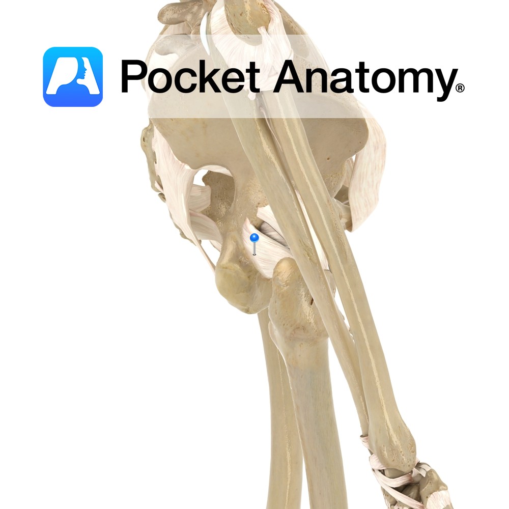
Origin:
Posterior gluteal line of the ilium, posterior aponeurosis of erector spinae, posterior surface of the sacrum , lateral aspect of the coccyx and the sacrotuberous ligament.
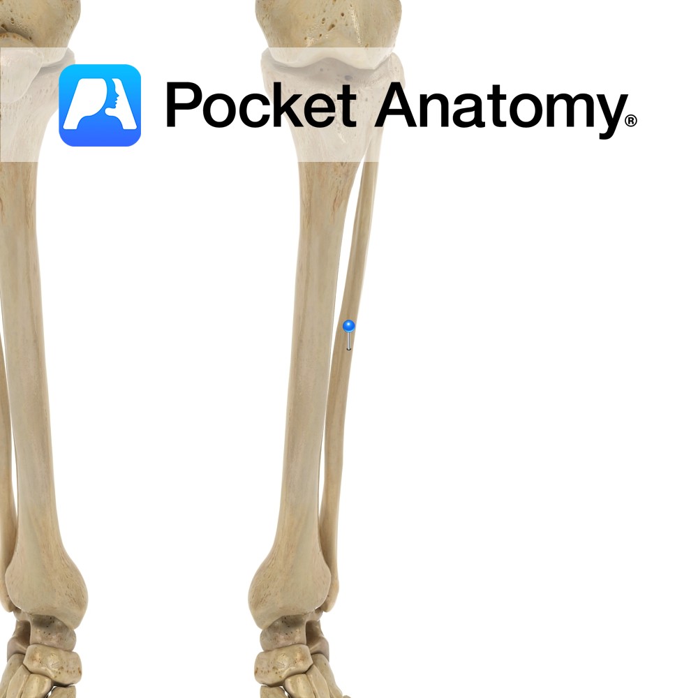
Insertion:
Iliotibial tract of the fascia lata and gluteal tuberosity of the femur.
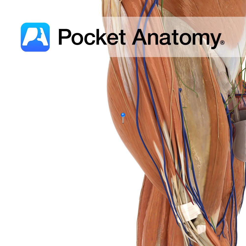
Key Relations:
The upper border of gluteus maximus lies anterior to the inferior third of gluteus medius.
Functions
-Extends the flexed thigh at the hip joint.
-Laterally rotates the hip joint.
-The upper fibres abduct the thigh at the hip joint.
-Stabilizes the hip joint to suppor t the trunk on the pelvis. (This action is what allowed us to develop bipedal gait.)
-Key in allowing the body to regain the erect postion after bending by rotating the pelvis backwards on the lower limbs.
-Also stabilizes the extended knee joint through the iliotibial tract.
Supply
Nerve Supply:
Inferior gluteal nerve (L5, S1, S2).
Blood Supply:
Branches of the internal iliac arteries including the superior and inferior gluteal arteries.
Clinical
The gluteal muscles are commonly used as a site for intramuscular injections. The needle is inserted into the upper and outer quadrant of the gluteal region to avoid the sciatic nerve. This is important as damage to the sciatic nerve can lead to paralysis of the hamstrings and all muscles below the knee, along with sensory loss of the skin below the knee (excluding the areas supplied by the saphenous nerve).
Interested in taking our award-winning Pocket Anatomy app for a test drive?
![]()