Anatomy
Origin:
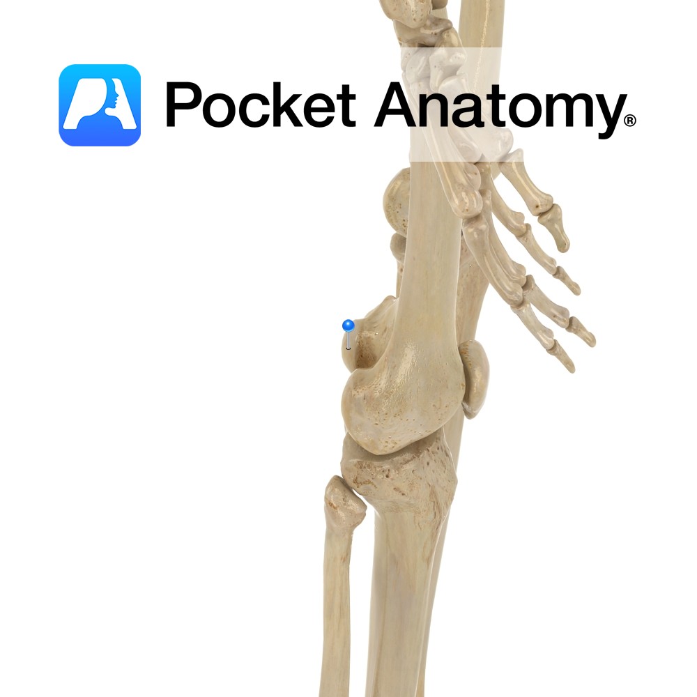
Outer surface of the ilium between the anterior and inferior gluteal lines.
Insertion:
The fibres converge to form a tendon that inserts on the anterior border of the greater trochanter of the femur.
Key Relations:
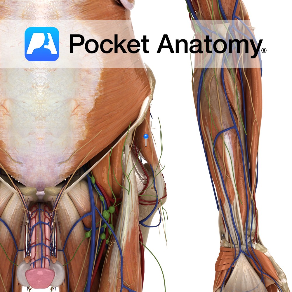
-Gluteus minimus lies posterior to gluteus medius.
-The deep branches of the superior gluteal vessels and the superior gluteal nerve are located between gluteus medius and minimus.
Functions
-Working with gluteus medius, it abducts and medially rotates the thigh at the hip joint.
-Both muscles are important in walking. When one leg is raised from the ground in walking both gluteus medius and minimus on the opposite side keep the trunk upright and prevent the pelvis from sagging on the unsupported moving side thereby stabilising the pelvis.
Supply
Nerve Supply:
Superior gluteal nerve (L4, L5, S1).
Blood Supply:
Branches of the internal iliac arteries including the superior and inferior gluteal arteries.
Clinical
Trendelenberg’s test is used to test hip stability. This involves asking the patient to stand unassisted on each leg, whilst the clinician places their fingers on the anterior superior iliac spines. Normally the hip is stabilized by gluteus medius and minimus. If the pelvis drops on the unsupported side this is a positive Trendelenberg’s sign. Thus may be caused by paralysis of gluteus medius and minimus, congenital dislocation of the hip, fracture of the neck of the femur or coxa vara.
Interested in taking our award-winning Pocket Anatomy app for a test drive?
![]()