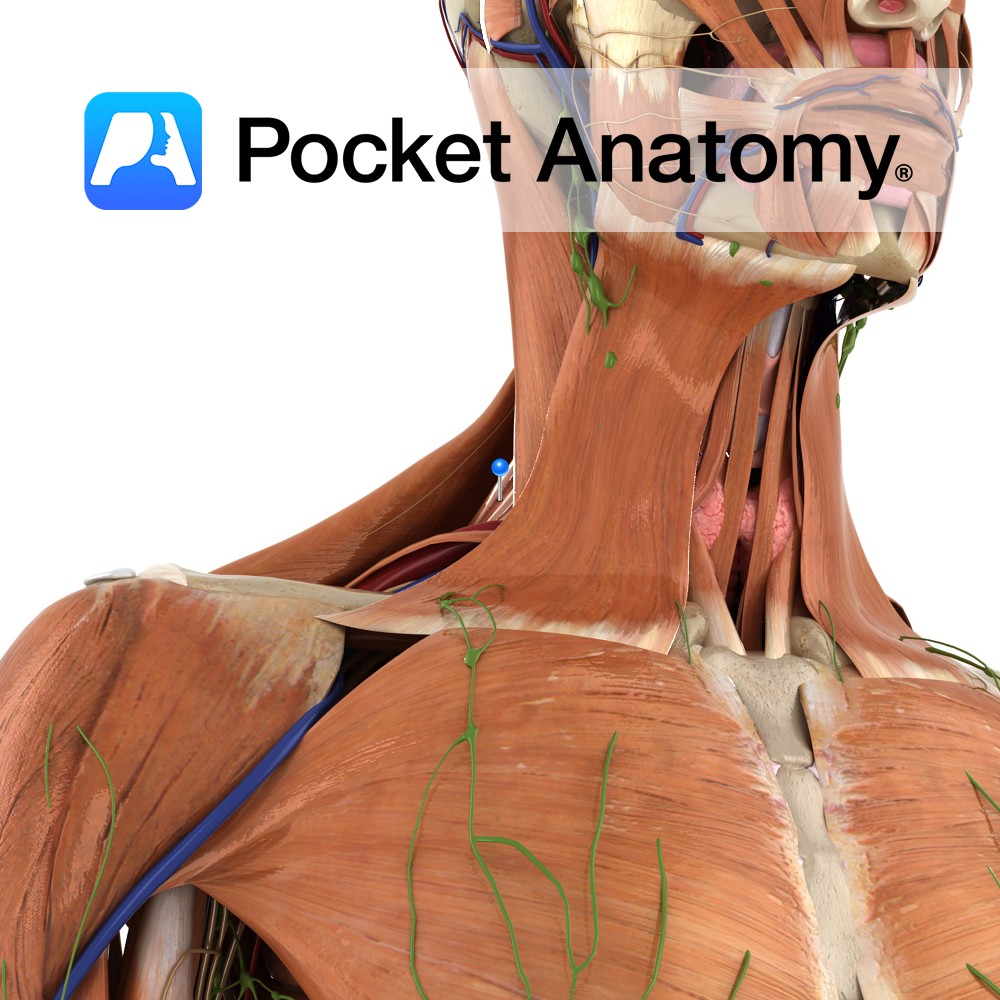
Anatomy
Origin:
Posterior tubercles of the transverse processes of the upper four cervical vertebrae (C1 to C4).
Insertion:
Medial border of the scapula between the superior angle and the spine, opposite the supraspinous fossa.
Key Relations:
Lies within the floor of the posterior triangle of the neck.
Functions
-Working with various other muscles of the upper limb, elevates e.g. shrugging shoulders or carrying a heavy bag, retracts and rotates the scapula. Elevation is its primary function.
-Also extends and laterally rotates the cervical spine.
Supply
Nerve Supply:
-Supplied directly by cervical nerves C3 and C4
-Dorsal scapular nerve (C5)
Blood Supply:
-Transverse and ascending cervical arteries
-Dorsal scapular artery.
Clinical
Levator scapulae syndrome: a soft tissue strain of the muscle most commonly referred to sports medicine practitioners or chiropractors.
Levator scapulae can be tested clinically similarly to the rhomboid muscles. The patient should be sitting upright with their arms flexed at the elbow and resting their hands in the small of their back. They should then try to externally rotate their arms against resistance from the clinician.
Interested in taking our award-winning Pocket Anatomy app for a test drive?
![]()

.jpg)