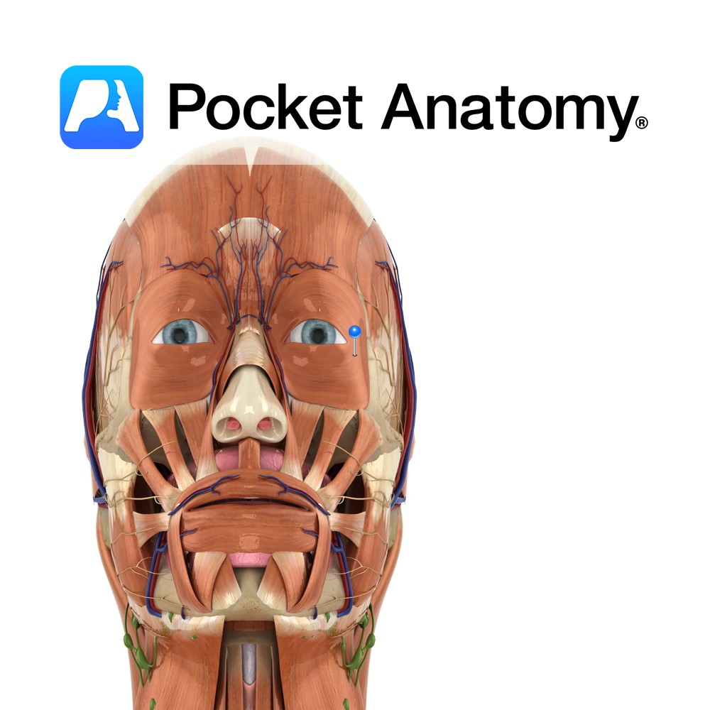
Anatomy
Origin:
Palebral part: Medial palpebral ligament.
Orbital part: Nasal portion of frontal bone, frontal process of maxilla, medial palebral ligament.
Insertion:
Palebral part: Lateral palebral raphe at outer corner of eye.
Orbital part: Fibres converge and form uninterrupted elipse around orbit of the eye.
Key Relations:
A third part of the muscle exists known as the lacrimal part however in some people it is very indistinct. It compresses the lacrimal sac and positions the lacrimal canals.
Functions
Closes the eyelids (only muscle in body capable of doing so).
Palebral part: Closes eyelids involuntarily and gently.
e.g. as in sleeping or blinking.
Orbital part: Closes eyelids voluntarily and forcibly.
e.g. when pretending to be asleep!.
Supply
Nerve Supply:
Temporal and zygomatic branches of the facial nerve (CN 7).
Blood Supply:
-Ophthalmic artery
–Superficial temporal artery
–Facial artery
-Infra-orbital branch of the maxillary artery.
Clinical
Lesions of the facial nerve such as those seen in Bell’s palsy result in an inability to blink or close the ipsilateral eyelid. The lack of irrigation of the eye that results increases the risk of corneal inflammation or ulceration.
Interested in taking our award-winning Pocket Anatomy app for a test drive?
![]()