Anatomy
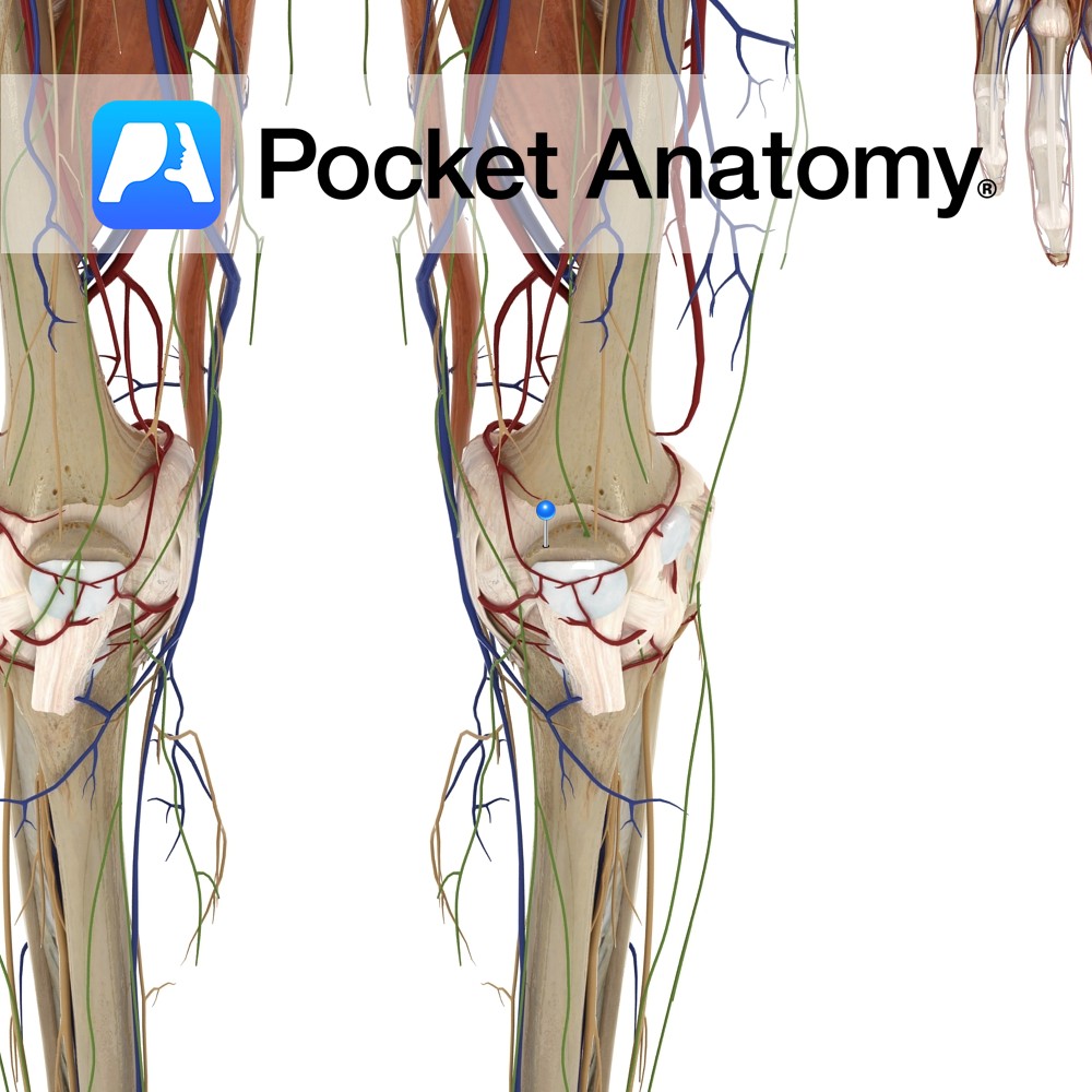
Kneecap, largest sesamoid (embedded in tendon) bone, triangular (base up, tip down) articulates with femur.
Clinical
Increases angle and leverage of quads in their extension of knee. Visible, palpable, moveable a little side to side when leg straight and quads relaxed. Patellar dislocation is usually lateral, accompanied by pain and swelling, can often be reduced (put back) when leg straightened, treated conservatively or surgically.
Vignette
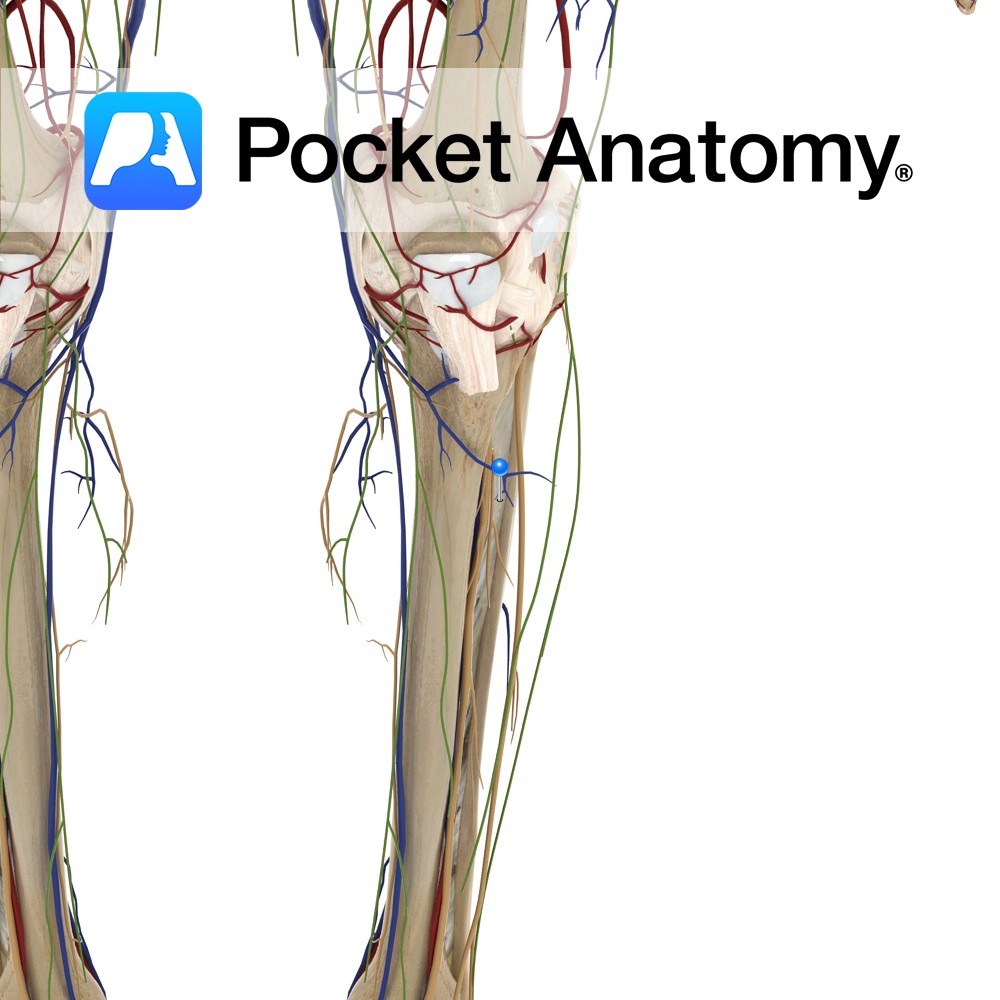
Many factors involved in stabilization of patella (ordinarily meaning prevented from lateral dislocation during quadriceps contraction); shape of articulating posterior surface (thicker in middle) which sits in the trochlear groove between the femoral condyles (lateral is thicker); vastus medialis insertion which pulls patella medially; tethering by medial and lateral retinaculae (parts of quads tendon either side patella that attach to tibia either side tuberosity); fibres of iliotibial tract.
Most stable at full extension.
Interested in taking our award-winning Pocket Anatomy app for a test drive?
![]()
.jpg)