Anatomy
Origin:
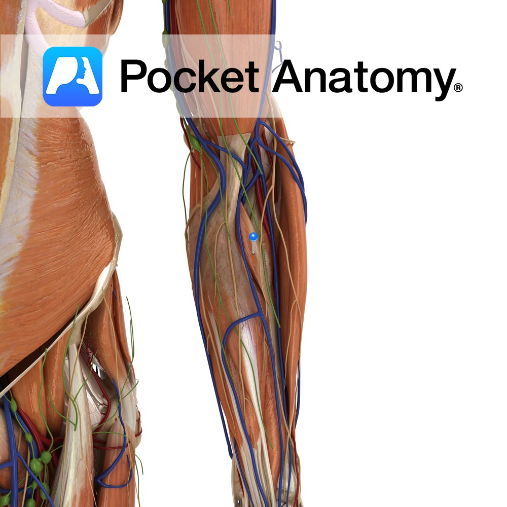
Humeral head: Medial epicondyle of humerus via the common flexor tendon, and adjacent supraepicondylar ridge.
Ulnar head: Medial aspect of the coronoid process of ulna.
Insertion:
Roughening on lateral surface of radial shaft near its midpoint.
Key Relations:
-The median nerve courses between the two heads of pronator teres in 80% of people.
-One of the four muscles in the superficial anterior compartment of the forearm.
-Forms the medial border of the cubital fossa.
Functions
-Pronation of forearm particularly when forceful or rapid pronation is required.
-Helps flex forearm at the elbow joint.
e.g. as in loosening a screw (right hand)..
Supply
Nerve Supply:
Median nerve (C6, C7).
Blood Supply:
–Ulnar artery
–Radial artery.
Clinical
Pronator syndrome is an entrapment neuropathy of the median nerve at the elbow as it passes between the heads of pronator teres. It is less common than carpal tunnel syndrome.
Interested in taking our award-winning Pocket Anatomy app for a test drive?
![]()