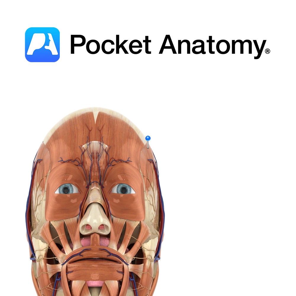
Anatomy
Origin:
Floor of temporal fossa and its overlying fascia.
Insertion:
Medial surface of coronoid process of mandible and anterior border of ramus of mandible.
Key Relations:
-Superficial to the muscle are the masseter and the temporal branches of the facial nerve.
-One of the four muscles of mastication.
Functions
-Elevates the mandible to occlude the teeth in mastication e.g. as in closing the mouth.
-Posterior fibres retract the mandible.
Supply
Nerve Supply:
Mandibular branch of the trigeminal nerve (CN 5).
Blood Supply:
Superficial temporal and deep temporal branches of the maxillary artery.
Clinical
Testing the bulk and power of the temporalis and masseter muscles can be useful in the detection of a CN 5 lesion. Muscle bulk can be palpated above the mandible. Power can be tested by asking the patient to bite forcefully onto a wooden tongue depressor and trying to remove it from their mouth. A bite of normal strength will prevent this.
Interested in taking our award-winning Pocket Anatomy app for a test drive?
![]()
.jpg)