Anatomy
Origin:
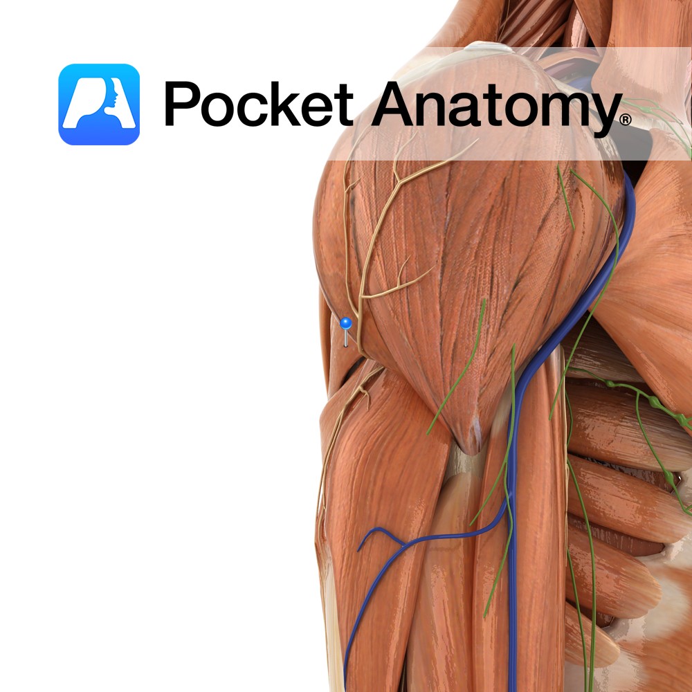
Oval area on posterior surface of inferior angle of scapula.
Insertion:
Medial lip of intertubercular sulcus on anterior surface of humerus.
Key Relations:
The teres minor and teres major muscles form the axillary space, through which several important arteries and veins pass.
Functions
-Medial rotation of the arm at the glenohumeral joint.
-Adduction of the humerus e.g. when tucking your shirt into the back of your trousers..
Supply
Nerve Supply:
Inferior subscapular nerve (C5, C6, C7).
Blood Supply:
-Thoracodorsal branch of subscapular artery
-Posterior circumflex humeral artery.
Clinical
Teres major is tested by asking the patient to abduct their arm to 90° and then to adduct the arm while resistance is applied from below the arm. The majority of the muscle can be felt contracting by feeling for the inferior angle of the scapula and then moving lateral to this along the posterior axillary fold.
Interested in taking our award-winning Pocket Anatomy app for a test drive?
![]()