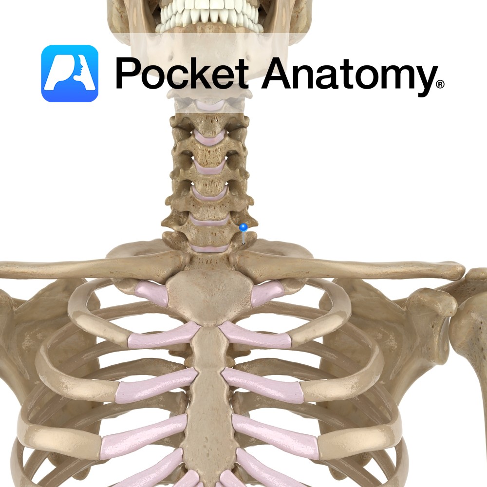
Anatomy
Has a heart-shaped body, lateral surface (each side) of which has an articulating facet for the head of 1st rib and a demi-facet for the upper half of the head of 2nd rib. From either side of the back of the body, pedicles project posteriorly, joining laminae which themselves meet at the midline (from where a spinous process projects back and down) to complete the neural arch (enclosing the vertebral foramen).
Laminae imbricate (overlap, tile-like) those of T2 below. Where pedicle meets lamina each side, a transverse process projects laterally (its tip articulates with tubercle of 1st rib) and a superior articular process projects up (for articulation with C7) and an inferior articular process projects down (for articulation with T2).
Clinical
Articulates with C7 above (top of vertebral body and 2 superior articular processes), T2 below (bottom of body and 2 inferior articular processes), 1st rib (costal facet on body and on transverse process) and 2nd rib (costal demi-facet on body).
The joints between upper and lower halves of heads of ribs and demi-facets of adjacent vertebral bodies are called zygapophyseal, and restrict flexion and extension, the main thoracic spinal movement being rotation.
Interested in taking our award-winning Pocket Anatomy app for a test drive?
![]()