
PocketAnatomy® is a registered brand name owned by © eMedia Interactive Ltd, 2009-2027.
iPhone, iPad, iPad Pro and Mac are trademarks of Apple Inc., registered in the U.S. and other countries. App Store is a service mark of Apple Inc.
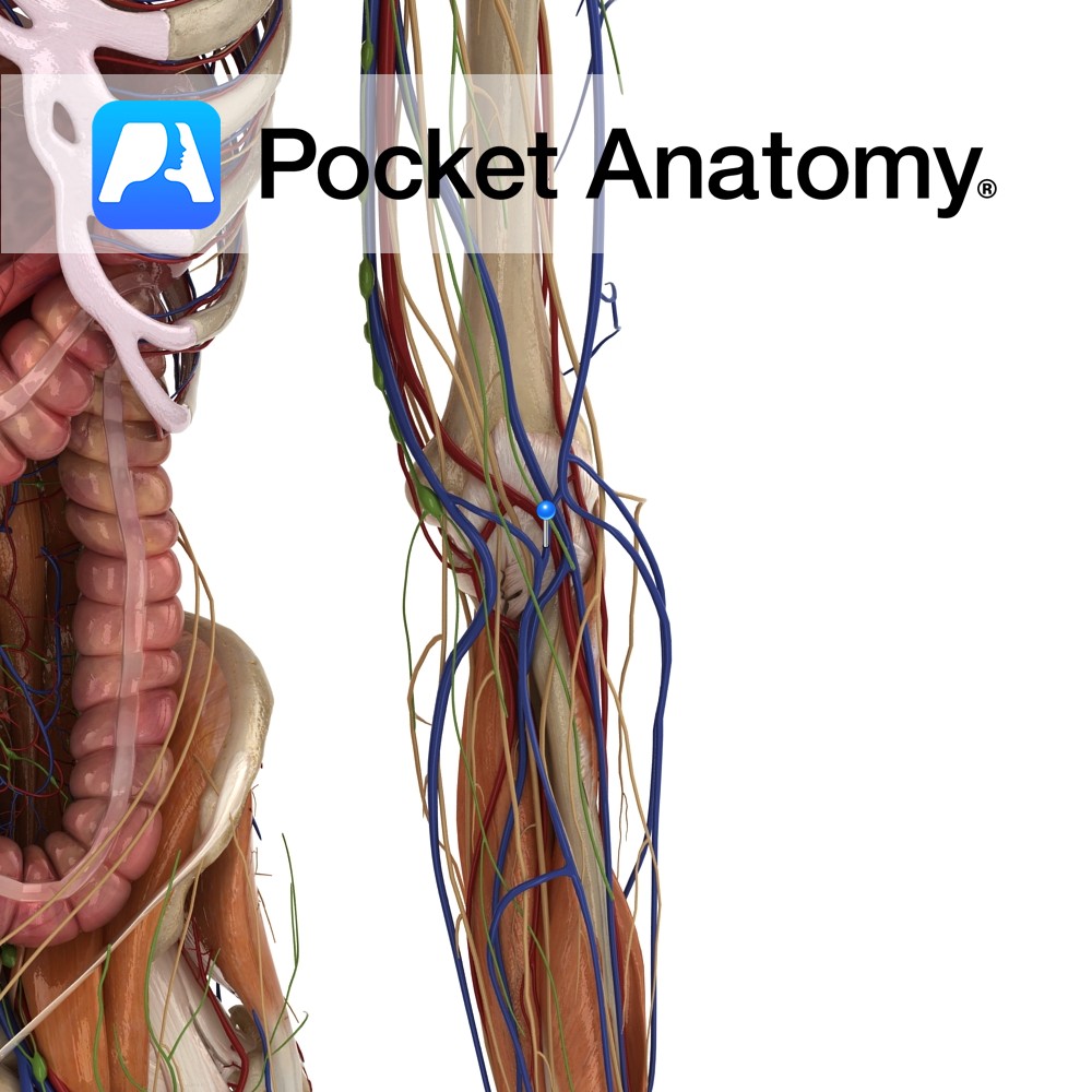
Anatomy Course A medium sized vein that comes off the antebrachial vein. At the elbow joint it joins the cephalic vein. Drain Drains the superficial component of the forearm. Clinical It is located significantly farther from arteries than other veins. Along with its size, it makes a popular target for inserting a cannula. Interested in
- Published in Pocket Anatomy Pins
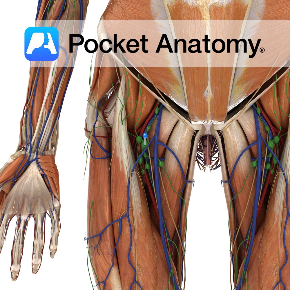
Anatomy Nodes in upper inner thigh, groin areas. Drain lymph from legs, outer/lateral aspect genitalia, lower abdominal wall. Interested in taking our award-winning Pocket Anatomy app for a test drive?
- Published in Pocket Anatomy Pins

Motion The intercarpal joints are synovial ellipsoid or spheroidal joints. The term intercarpal joints refers to the joints between the proximal row of carpal bones, the joints between the distal row of carpal bones and the complex midcarpal joint between the two rows. The movements of the intercarpal joints can be considered with those of
- Published in Pocket Anatomy Pins

Anatomy Course Anterior division of the first thoracic spinal nerve. Runs along the first intercostal space, below the superior rib until it ends as the anterior cutaneous branch which surfaces to supply the cutaneous skin. Rarely is a lateral cutaneous branch associated with this nerve. Supply Somatic motor innervation to the thoracic wall muscles, sensory
- Published in Pocket Anatomy Pins
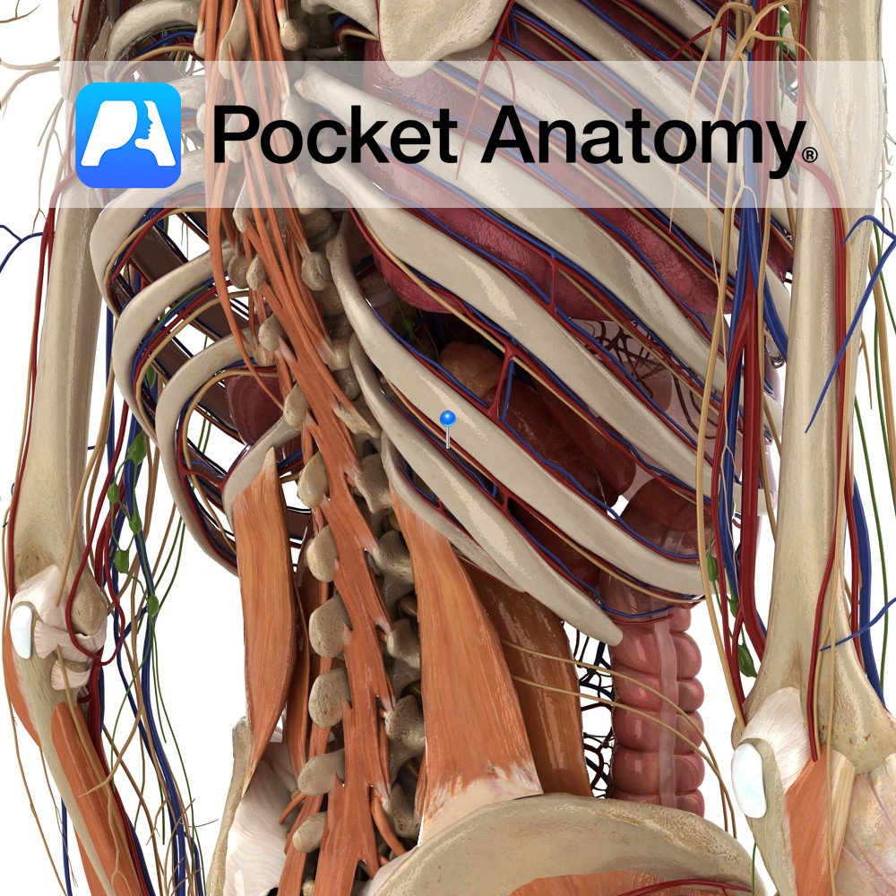
Anatomy Course Anterior division of the tenth thoracic spinal nerve. Runs along the tenth intercostal space, below the superior rib until it ends as the anterior cutaneous branch which surfaces to supply the cutaneous skin. Along the way it gives off a lateral cutaneous branch that emerges superficially and splits into a posterior and anterior
- Published in Pocket Anatomy Pins
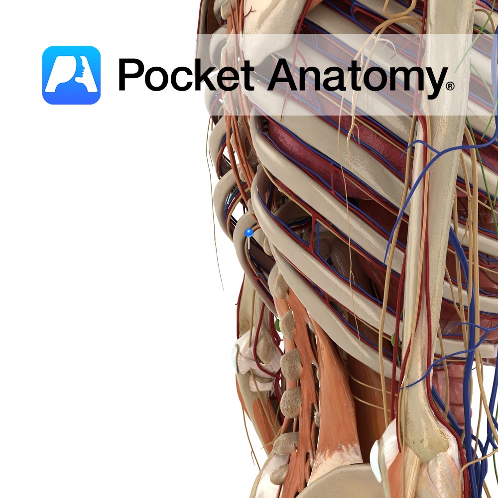
Anatomy Course Anterior division of the eleventh thoracic spinal nerve. Runs along the eleventh intercostal space, below the superior rib until it ends as the anterior cutaneous branch which surfaces to supply the cutaneous skin. Along the way it gives off a lateral cutaneous branch that emerges superficially and splits into a posterior and anterior
- Published in Pocket Anatomy Pins

Anatomy Course Anterior division of the second thoracic spinal nerve. Runs along the second intercostal space, below the superior rib until it ends as the anterior cutaneous branch which surfaces to supply the cutaneous skin. Along the way it gives off a lateral cutaneous branch that emerges superficially and splits into a posterior and anterior
- Published in Pocket Anatomy Pins
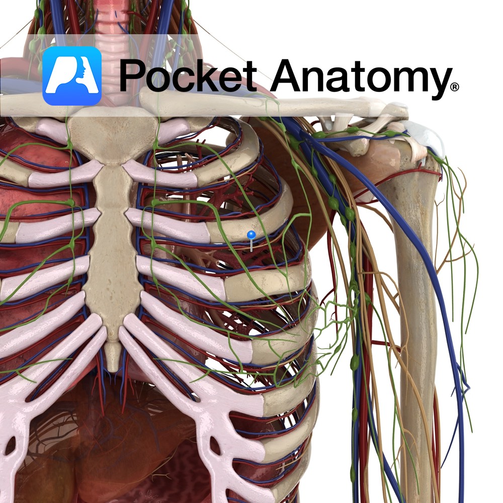
Anatomy Course Anterior division of the third thoracic spinal nerve. Runs along the third intercostal space, below the superior rib until it ends as the anterior cutaneous branch which surfaces to supply the cutaneous skin. Along the way it gives off a lateral cutaneous branch that emerges superficially and splits into a posterior and anterior
- Published in Pocket Anatomy Pins
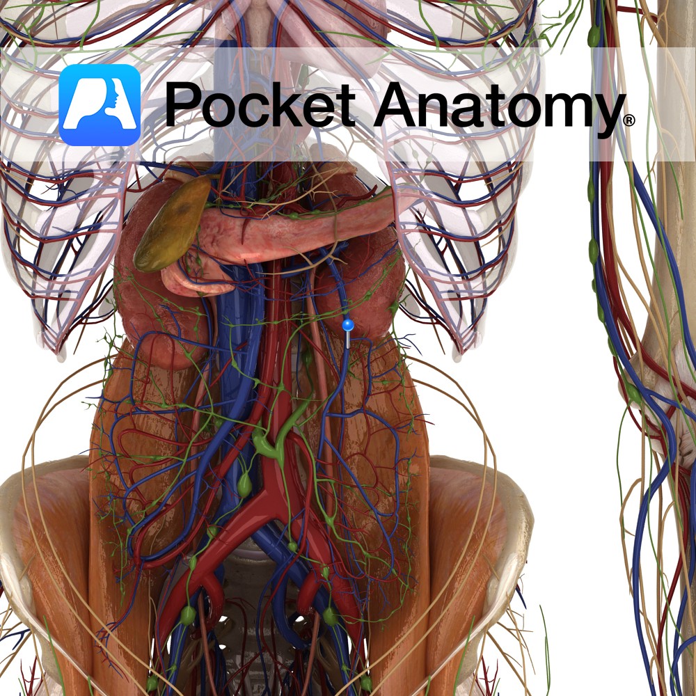
Anatomy Course Receives blood rich in nutrients from the large intestine via its tributaries and then terminates by joining with the splenic vein to form the portal vein. Drain Drains the large intestine, sigmoid colon and the rectum. Interested in taking our award-winning Pocket Anatomy app for a test drive?
- Published in Pocket Anatomy Pins

Anatomy Paired. Turbinated. Forms inferior meatus at lower part of lateral wall of nasal cavity, presents big surface area for air-conditioning (filter, warm, moisten). Is covered by mucous membrane and perforated by nasolacrimal duct. Articulates with ethmoid, maxilla, lacrimal and palatine. Clinical Superior nasal concha not a separate bone; part of ethmoid. Vignette Concha (Spanish);
- Published in Pocket Anatomy Pins