
PocketAnatomy® is a registered brand name owned by © eMedia Interactive Ltd, 2009-2027.
iPhone, iPad, iPad Pro and Mac are trademarks of Apple Inc., registered in the U.S. and other countries. App Store is a service mark of Apple Inc.
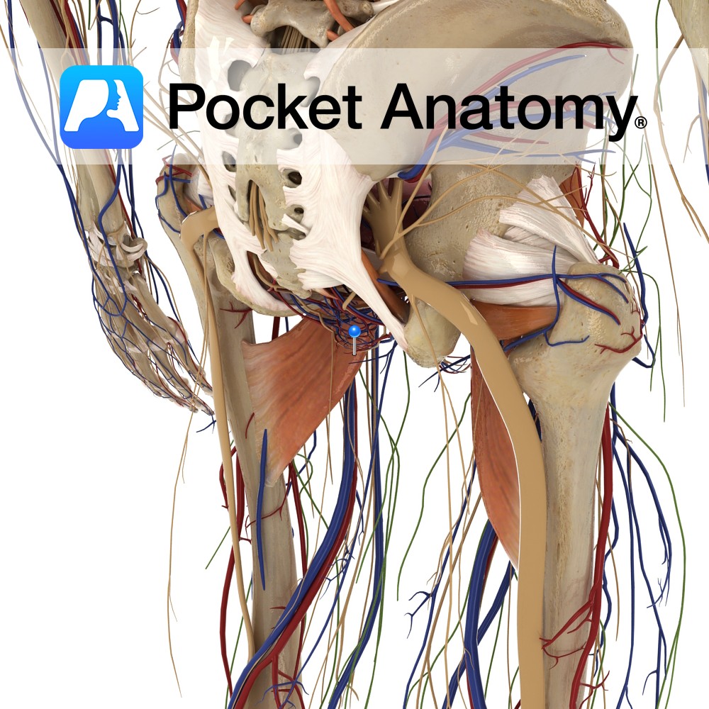
Anatomy Course Branches off the internal pudendal artery as it crosses the ischial tuberosity. It promptly pierces the pudendal cavity and gives off its branches. Supply Responsible for supplying the lower half of the rectum as well as some branches to the superficial gluteal region. Interested in taking our award-winning Pocket Anatomy app for a
- Published in Pocket Anatomy Pins
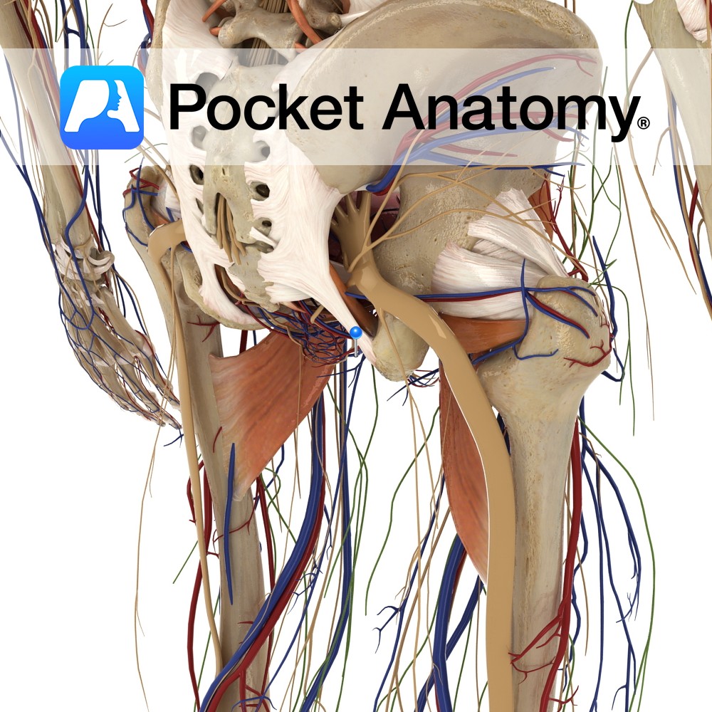
Anatomy Course Begins at the inferior border of the hemorrhoidal plexus where it travels shortly to empty into the internal pudendal vein. Drain Drains the inferior rectum. Interested in taking our award-winning Pocket Anatomy app for a test drive?
- Published in Pocket Anatomy Pins
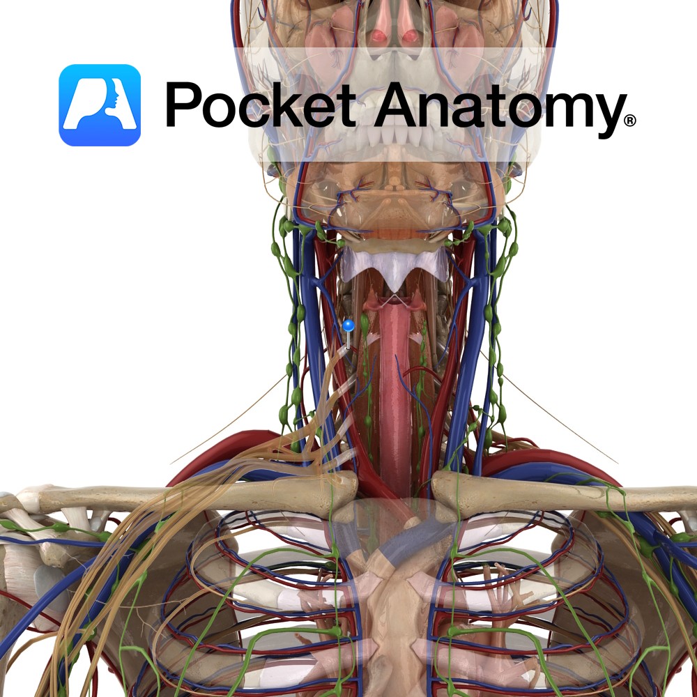
Anatomy Course Branch of the thyrocervical trunk of the subclavian artery. As it rises, it passes behind the carotid sheath and the sympathetic trunk. The middle cervical ganglion rests on the artery. Supply Supplies the lower part of the thyroid gland as well as the parathyroid glands. Interested in taking our award-winning Pocket Anatomy app
- Published in Pocket Anatomy Pins
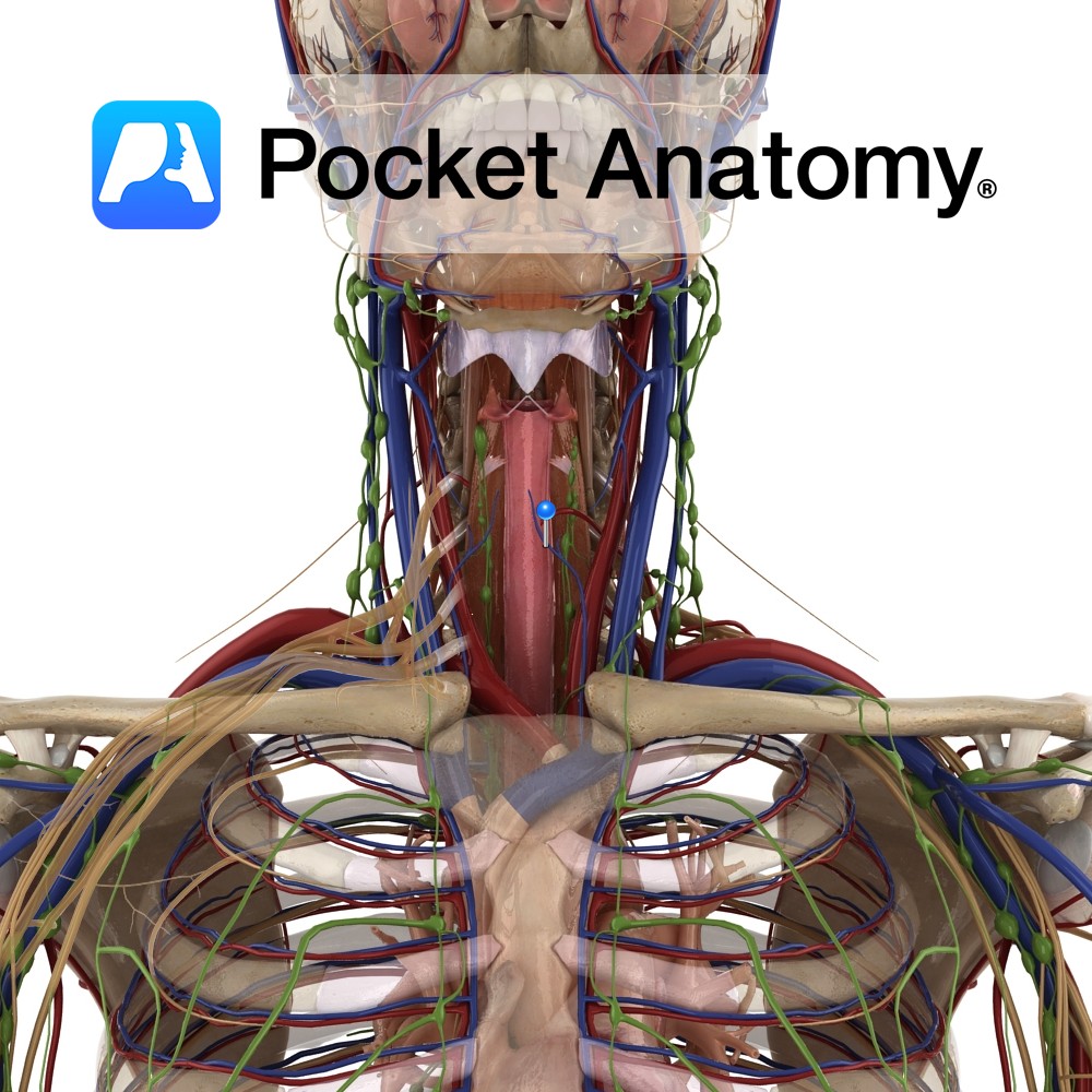
Anatomy Course Commences in the thyroid venous plexus that is located in front of the trachea. Usually divides into several veins. The right veins descend across the innominate artery and drain into the right innominate artery. The left veins empty into the left innominate trunk. Drain Drains structures of the neck such as the trachea,
- Published in Pocket Anatomy Pins
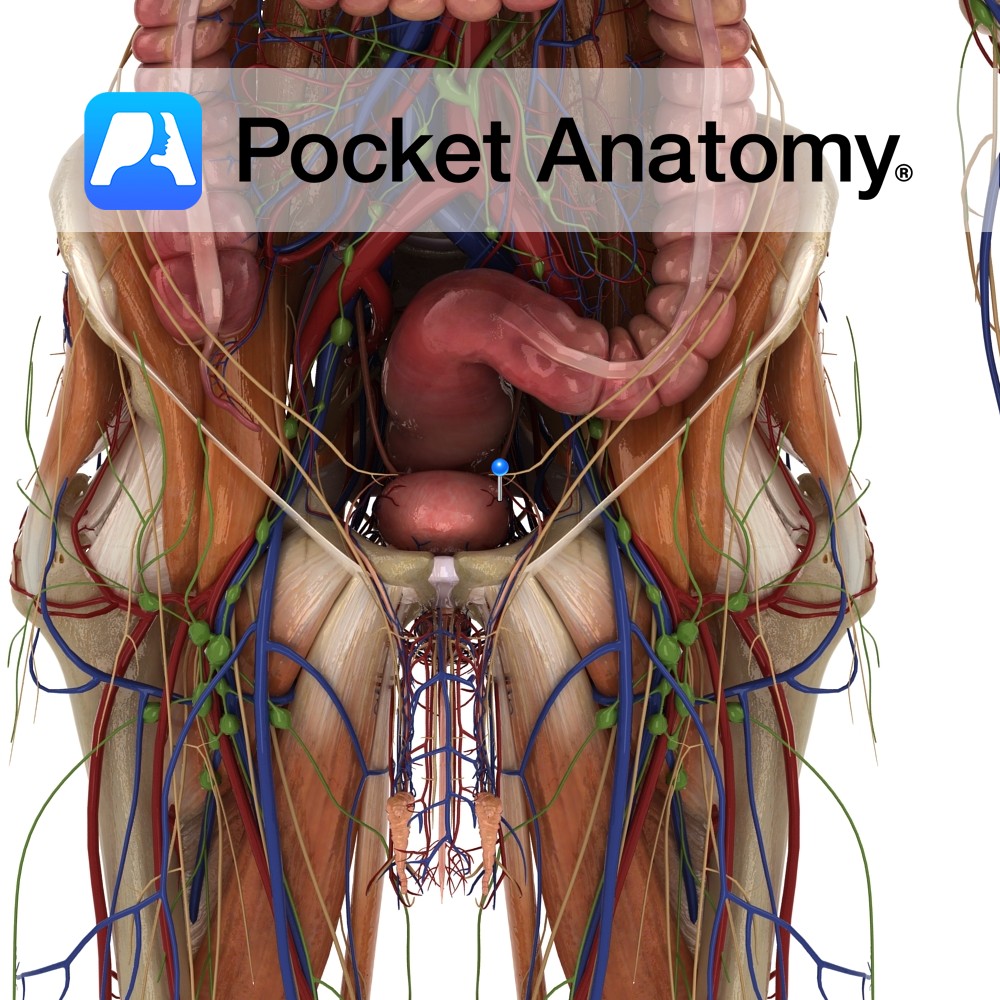
Anatomy Course Usually arises as a branch of the internal iliac artery but sometimes it may arise from the middle rectal artery. Supply Supplies the bladder, prostate, ureters and seminal vesicles. Interested in taking our award-winning Pocket Anatomy app for a test drive?
- Published in Pocket Anatomy Pins
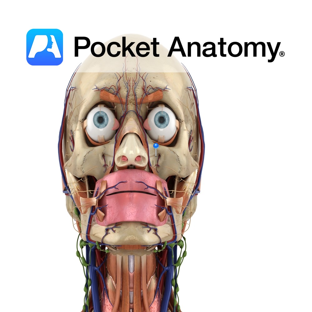
Anatomy Course The maxillary nerve (V2) is one of the branches of the trigeminal nerve (V). It branches from the trigeminal nerve whilst it is still in the middle cranial fossa and then exits the inside of the skull through the foramen rotundum. From here, it crosses the pterygopalatine fossa and enters the eye socket.
- Published in Pocket Anatomy Pins
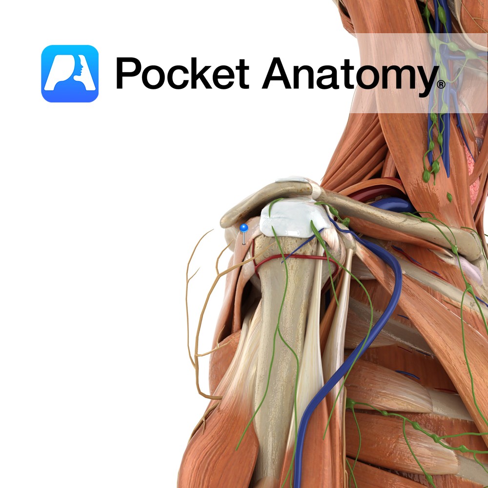
Anatomy Origin: Medial two-thirds of infraspinous fossa of scapula and infraspinous fascia that covers the muscle. Insertion: Middle facet of greater tubercle of humerus. Key Relations: -The tendon of infraspinatus is sometimes separated from the capsule of the shoulder joint by a bursa, which may communicate with the joint cavity. -One of the four muscles
- Published in Pocket Anatomy Pins
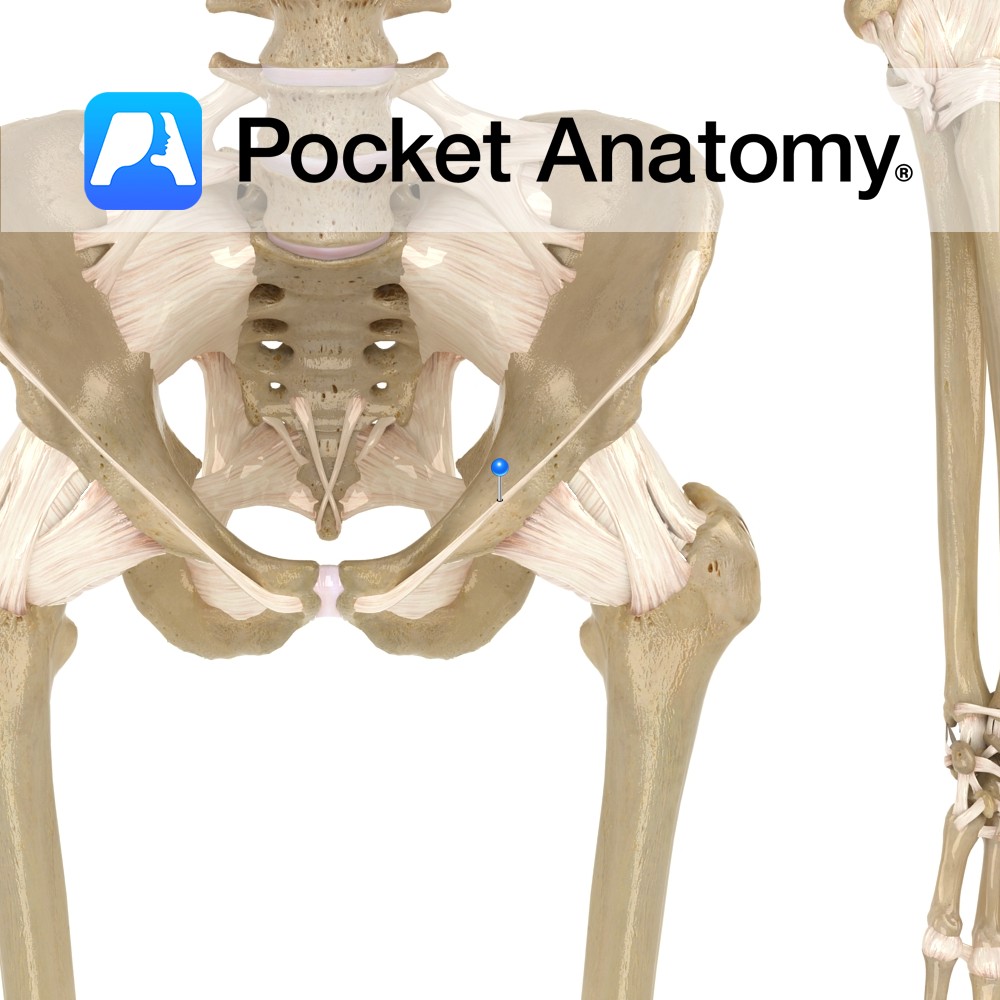
Anatomy A thickening of the lower border of the abdominal oblique muscles, attaching from the anterior superior iliac spine to the pubic tubercle medially. Functions Forms the inguinal canal and part of the tendon of the external obliques. Interested in taking our award-winning Pocket Anatomy app for a test drive?
- Published in Pocket Anatomy Pins
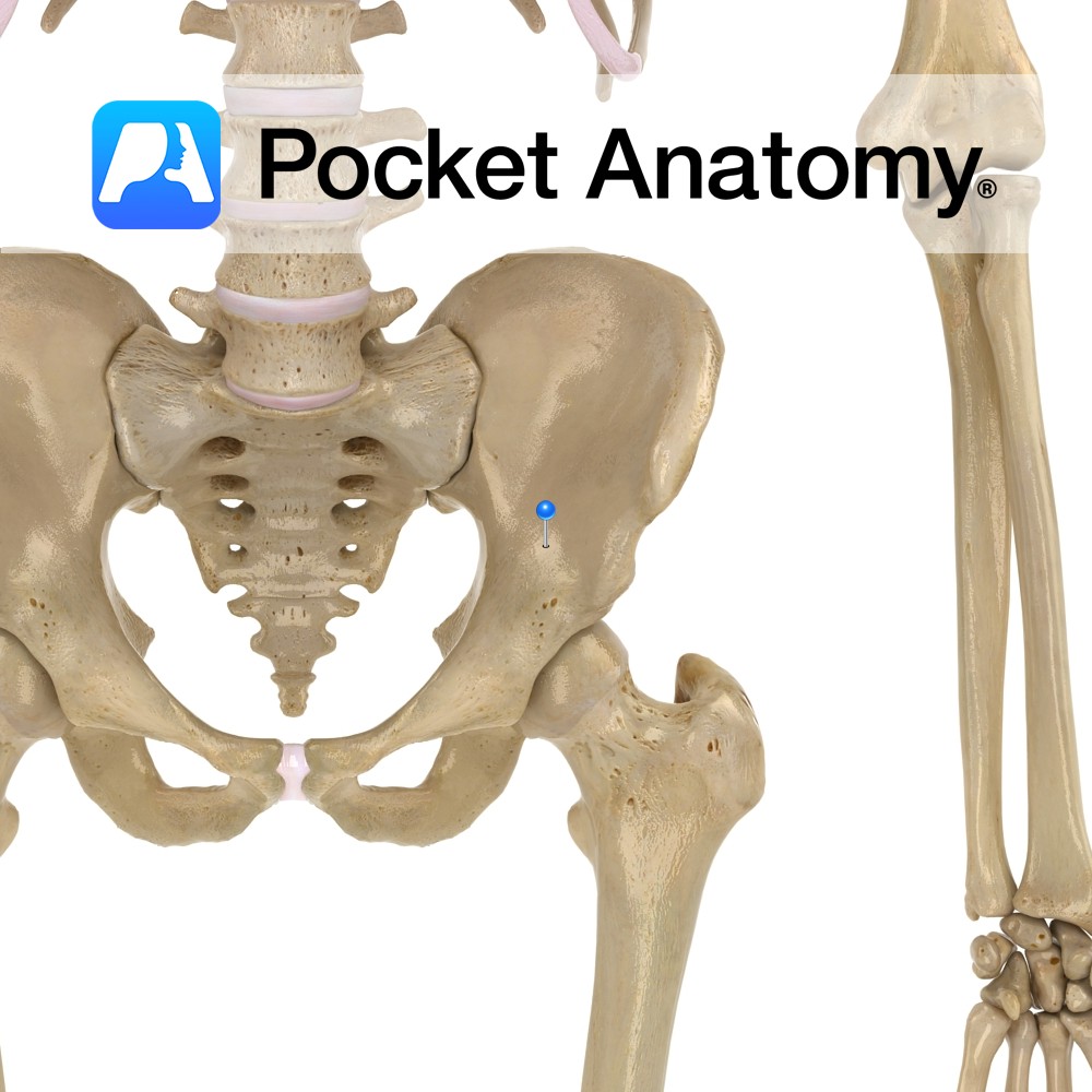
Anatomy The pelvic brim (edge of pelvic inlet) is formed by the arcuate lines of the ilia, the pectineal lines of the pubis, the upper margin of the pubic symphysis and the sacral promontory. Above, abdominal cavity; below, pelvic cavity. Vignette Viewed from above, the pelvic brim is smooth, almost circular. Interested in taking our
- Published in Pocket Anatomy Pins
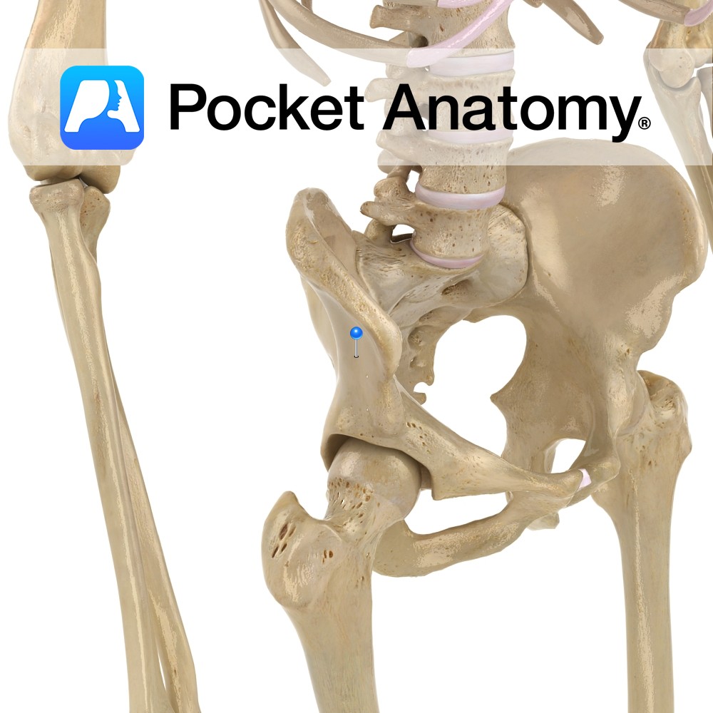
Anatomy The outside (lateral) surface of the wing (ala) of the ilium, site for attachment (from back to front) of gluteus maximus, medius, minimus. Interested in taking our award-winning Pocket Anatomy app for a test drive?
- Published in Pocket Anatomy Pins