PocketAnatomy® is a registered brand name owned by © eMedia Interactive Ltd, 2009-2022.
iPhone, iPad, iPad Pro and Mac are trademarks of Apple Inc., registered in the U.S. and other countries. App Store is a service mark of Apple Inc.

Ala of Sacrum anatomy Ala of sacrum is a large triangular surface either side of sacral base, continuous with iliac fossa (akin to adapted and joined transverse and costal processes elsewhere spine). Interested in taking our award-winning Pocket Anatomy for a test drive?
- Published in Pocket Anatomy Pins, Teaching Anatomy
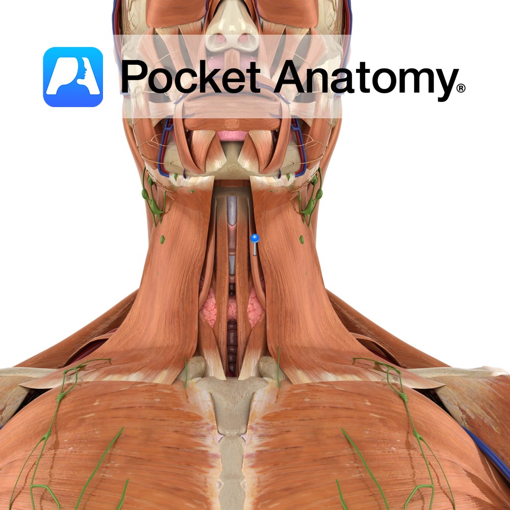
Anatomy Origin: Superior border of scapula medial to suprascapular notch. Insertion: Body of hyoid bone. Key Relations: -Is one of the infrahyoid muscles (sometimes referred to as ‘strap muscles’) lying in the muscular triangle of the neck. -Lateral to sternohyoid. Functions Depresses and fixes the hyoid bone. Supply Nerve Supply: Anterior rami of C1 to
- Published in Pocket Anatomy Pins, Teaching Anatomy
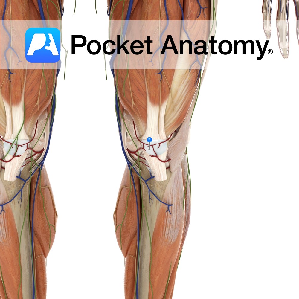
Anatomy Situated in front of the patella bone. Functions Acts as a cushion between bone and skin reducing friction between both. Clinical Prepatellar bursitis or ‘housemaid’s knee’ is a common injury to the bursa. The bursa becomes swollen due to repetitive work in a kneeling position. Interested in taking our award-winning Pocket Anatomy app for
- Published in Pocket Anatomy Pins, Teaching Anatomy
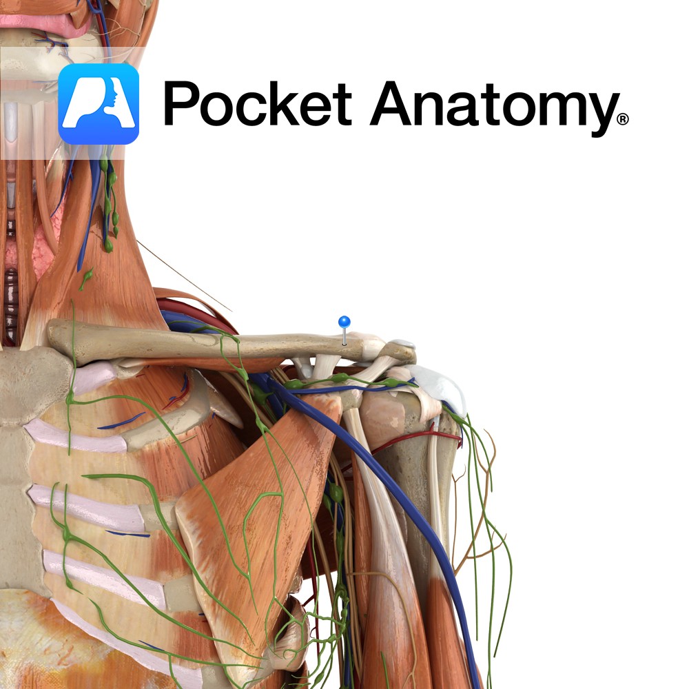
Motion The acromioclavicular joint is classified as a multiaxial synovial plane joint but it is more helpful to think of it as a pivot point. As the name suggests it is the articulation of the acromion of the scapula with the clavicle. It allows you to raise your arms above your head i.e. scapular elevation
- Published in Pocket Anatomy Pins, Teaching Anatomy
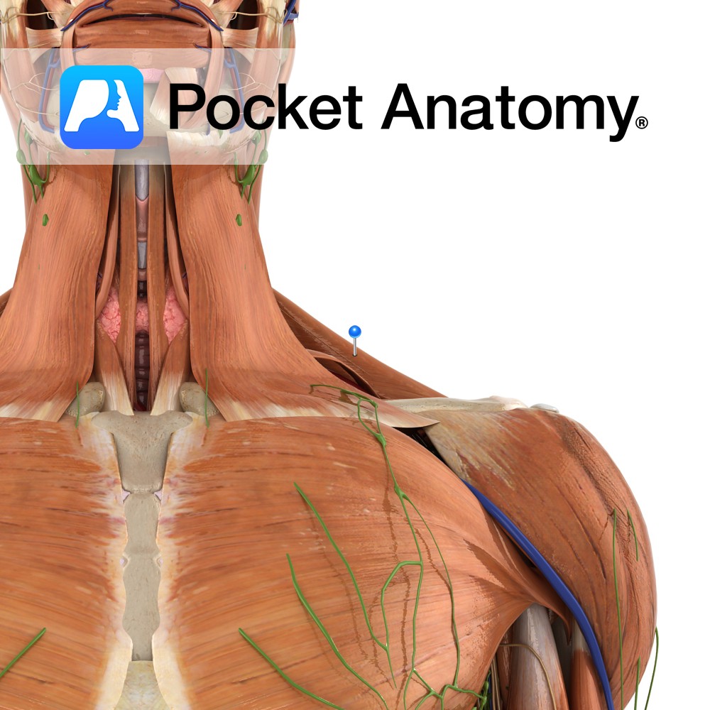
Anatomy Origin: Superior nuchal line of the occipital bone, external occipital protuberance, ligamentum nuchae and spinous processes of C7 to T12. Insertion: Superior fibres: posterior border of the lateral third of the clavicle. Middle fibres: medial border of the acromion and the superior edge of the spine of the scapula. Inferior fibres: tubercle of the
- Published in Pocket Anatomy Pins, Teaching Anatomy
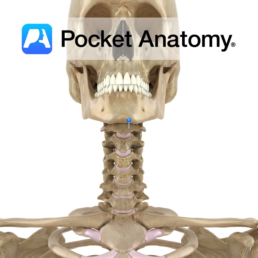
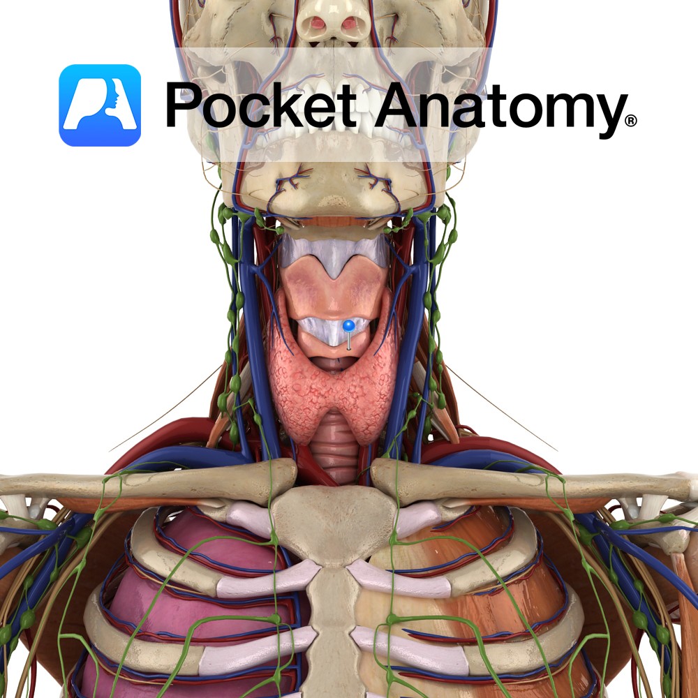
Anatomy Lingual bone, U-shaped, in front of neck, between larynx (behind) and mandible (in front), at level of C3; made up of body (transverse), greater horns/cornus (long, pointing back), lesser cornus/horns (short, pointing up); suspended from tips of styloid processes of mandible by ligaments (this syndesmotic connection being its only bony articulation); anchored by multiple
- Published in Pocket Anatomy Pins, Teaching Anatomy
Anatomy The most inferior part of the laryngeal cartilages and completely encircles the trachea. It is shaped like a signet ring with a broad lamina of cricoid cartilage posteriorly and a much slimmer arch of cartilage anteriorly completing the circle. The lamina has two depressions separated by a vertical ridge. There are two articular facets
- Published in Pocket Anatomy Pins, Teaching Anatomy

Health Literacy: when I was caring for patients in clinical practice, I spent time teaching them about their bodies, the conditions that brought them for care, and reviewing the results of any laboratory tests that had been done, as well as discussing the medications that had been prescribed. In each of these situations, I relied
- Published in Main Blog, Teaching Anatomy

Learning Anatomy: the human body is an exciting frontier to explore—from the first time you outline your body on paper in kindergarten to see how all the parts fit together; to middle school when you learn how stomach acids work to digest food; to high school when you’ve broken your arm playing football; to college
- Published in Main Blog, Teaching Anatomy
- 1
- 2