Anatomy
Origin:
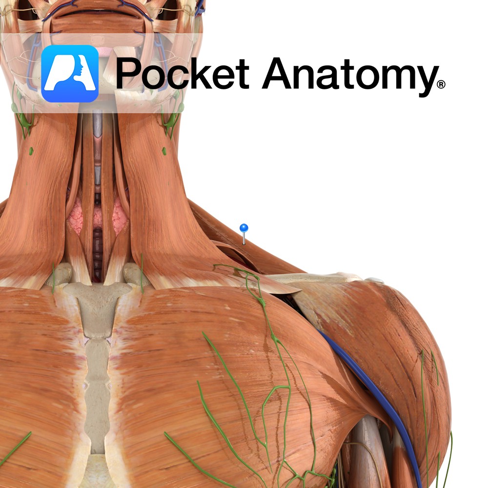
Superior nuchal line of the occipital bone, external occipital protuberance, ligamentum nuchae and spinous processes of C7 to T12.
Insertion:
Superior fibres: posterior border of the lateral third of the clavicle.
Middle fibres: medial border of the acromion and the superior edge of the spine of the scapula.
Inferior fibres: tubercle of the spine of the scapula.
Key Relations:
Trapezius forms one of the boundaries of the posterior triangle of the neck and the superiomedial border of the triangle of auscultation.
Functions
-Superior fibres elevate the scapula e.g. shrugging the shoulders.
-Middle fibres retract the scapula.
-Inferior fibres depress the scapula.
-The superior and inferior fibres work together to rotate the scapula e.g. overhead movements..
Supply
Nerve Supply:
–Accessory nerve (CN11)
-Anterior rami of cervical nerves C3 and C4
Blood Supply:
-Transverse cervical artery
-Dorsal scapular artery.
Clinical
Accessory nerve palsy can result in a palsy of trapezius and drooping of the arm at the shoulder may be observed.
The function of the trapezius muscle can be tested clinically by asking patients to shrug their shoulders against resistance.
Interested in taking our award-winning Pocket Anatomy app for a test drive?
![]()
-bone.jpg)