Anatomy
Origin:
Body and inferior ramus of pubic bone.
Insertion:
Pectineal line and proximal part of linea aspera of the femur.
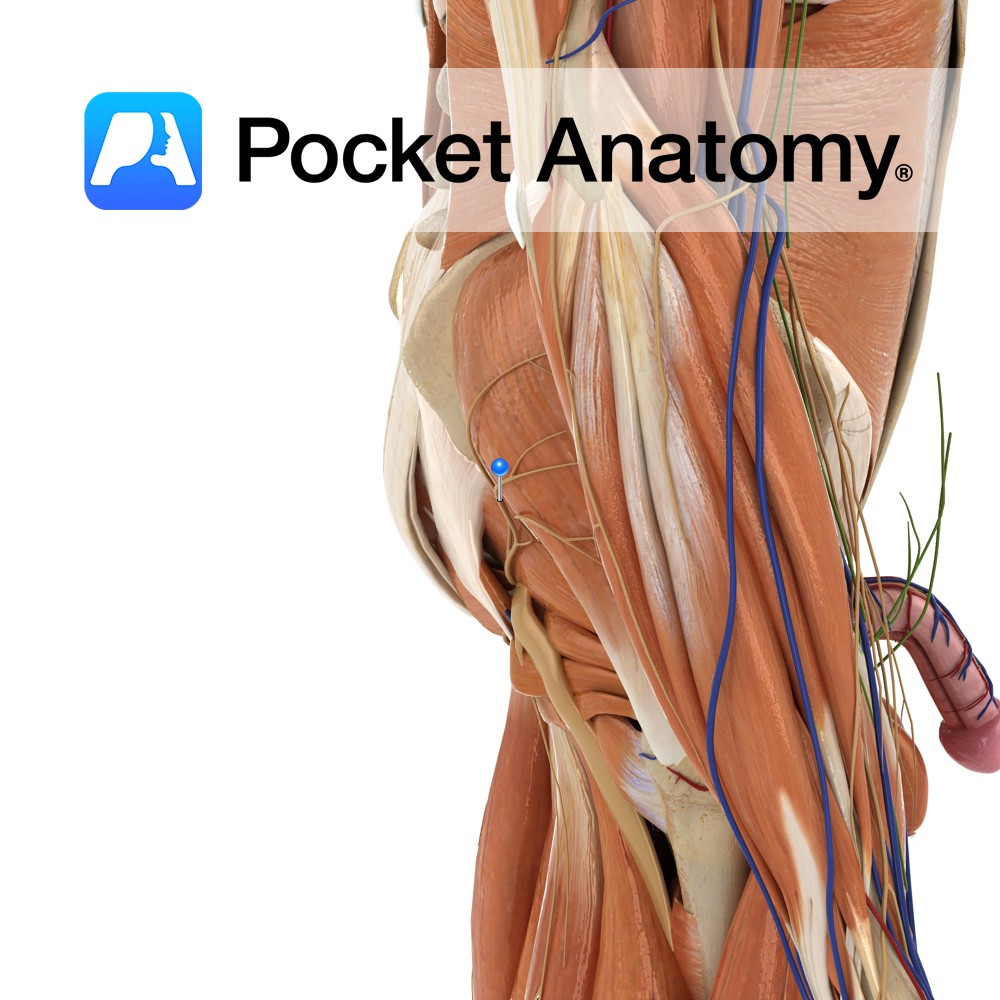
Key Relations:
-One of the six muscles of the medial compartment of the thigh.
-Lies posterior to pectineus, adductor longus and the anterior branch of the obturator nerve.
-Posterior to adductor brevis are adductor magnus and the posterior branch of the obturator nerve.
-Its upper border is associated with the medial circumflex femoral artery.
-Near its femoral insertion the muscle is pierced by the second or first and second perforating arteries (branches of the deep artery of the thigh).
Functions
Adducts the thigh at the hip joint.
Supply
Nerve Supply:
Obturator nerve (L2, L3, L4).
Blood Supply:
–Obturator artery
-Medial circumflex femoral artery.
Clinical
Hip adductor injuries (groin strains) are common sporting injuries and may involve any one of the adductor muscles of the thigh (pectineus, adductor brevis, adductor longus, gracillis, adductor magnus). Damage can occur when high demands are placed upon the muscles such as changing direction rapidly. Another common cause observed with soccer players is forced abduction of the thigh during an intended adduction. This scenario can occur when the player trying to kick the ball in one direction meets resistance, such as a player attempting to kick it in the opposite direction.
Treatment is generally non-operative. Rest and anti-inflammatory medication are used early on, followed by increasing exercise and range of motion work.
The adductor muscles of the thigh are tested as a functional group. The patient is requested to lie with their knees extended on the opposite side of that to be tested. The physician will abduct the leg on the side being tested, and then ask the patient to adduct the leg against resistance.
Interested in taking our award-winning Pocket Anatomy app for a test drive?
![]()