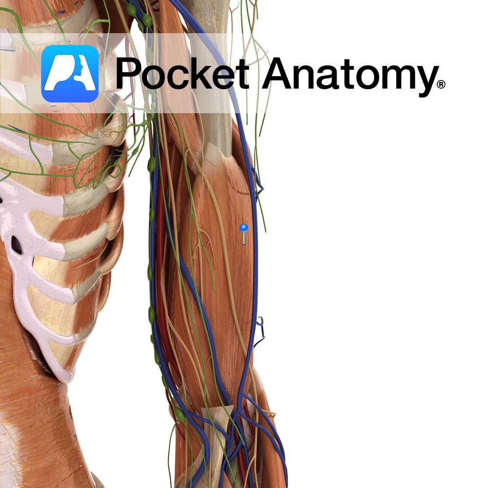
Anatomy
Origin:
Distal half of anterior surface of the humerus.
Insertion:
Coronoid process and the tuberosity of the ulna.
Key Relations:
-Located deep to the biceps brachii and forms part of the floor of the cubital fossa.
–Musculocutaneous nerve lies on its lateral surface adjacent to biceps brachii.
Functions
Works synergistically with biceps brachii for flexion of the forearm on the arm at the elbow joint. Is the most powerful flexor of the forearm.
Supply
Nerve Supply:
Musculocutaneous nerve (C5, C6).
Blood Supply:
–Radial recurrent artery
–Brachial artery.
Clinical
Brachialis is tested clinically by passive and active (against resistance) flexion of the forearm at the elbow, i.e. ask patient to bend arm at elbow and then bring arm towards the shoulder as the clinician attempts to push arm downwards away from shoulder.
Interested in taking our award-winning Pocket Anatomy app for a test drive?
![]()