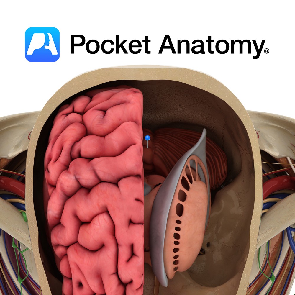
Anatomy
Located in the midline, between the two cerebellar hemispheres. It is further subdivided into nine areas, from anterior to posterior, as follows: lingula, central lobule, culmen, declive, folium, tuber, pyramid, uvula and nodule.
Blood Supply:
Supplied by branches of the superior, anterior inferior and posterior inferior cerebellar arteries.
Clinical
Cerebellar vermis lesions are characterised by axial dysequilibrium, broad-based ataxic gait, with minimal or no appendicular (limb) ataxia, and occasionally presence of spontaneous nystagmus (if caudal vermis involved). Chronic alcohol toxicity commonly displays localised pathology to the rostral vermis. Medulloblatomas may preferentially involve the caudal vermis.
Dandy-Walker syndrome is a rare congenital anomaly, which involves complete or partial absence of the vermis, and large fourth ventricle cisterna magna cyst. Although there is absence of the vermis, ataxia is unusual, and any clinical signs often arise from associated defects e.g. hydrocephalus (which occurs in approximately 85% of cases of Dandy-Walker).
Interested in taking our award-winning Pocket Anatomy app for a test drive?
![]()