Anatomy
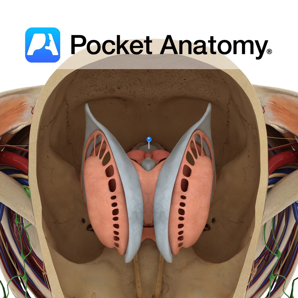
Is a rhomboid or diamond shaped cavity lined with ependymal cells part of the series of fluid-filled cavities, which make up the ventricular system. It is located anterior to the cerebellum and posterior to the pons and superior half of the medulla. It is connected superiorly to the third ventricle via the cerebral aqueduct of Sylvius.
The roof is formed by the cerebellum. The floor is formed by the rhomboid fossa. It is continuous inferiorly with the central canal of the spinal cord.
The fourth ventricle has 3 openings into the subarachnoid space: the median aperture known as the foramen of Magendie and the 2 lateral apertures known as the foramina of Luschka.
Functions
Filled with cerebrospinal fluid (CSF) and drains CSF from the third ventricle to the subarachnoid space.
Clinical
Hydrocephalus denotes ventricular enlargement. Hydrocephalus may be communicating (i.e not obstructive) e.g. normal pressure hydrocephalus, or non-communicating (i.e. obstructive hydrocephalus) e.g. colloid cyst obstructing the third ventricle. Normal pressure hydrocephalus is associated with gait impairment, cognitive impairment, and urinary incontinence.
Interested in taking our award-winning Pocket Anatomy app for a test drive?
![]()


.jpg)