Anatomy
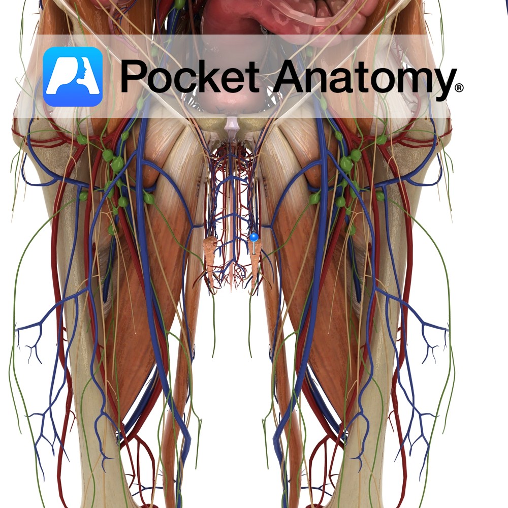
Epididymis (head – receives spermatozoa, body, tail – reabsorbs fluids and concentrates sperm) is a single coiled tube, 20-24 feet long, enveloping/investing/covering the back and top of the testis, its head continuous with and receiving immature sperm (can’t swim or fertilise) from efferent ducts at back of testis, passing them on (mature, typically after 2 months) during ejaculation to the vas/ductus deferens.
Aberrant Ductules: Outpouchings (diverticuli) of the epididymis; superior (head) and inferior (tail) aberrant ductules. They are vestigial remnants (also paradidymis) of embryonic mesonephric (Wolffian) ducts (which develop into vas deferens and head of epididymis).
Physiology
A reservoir, maturation site and concentrator (particularly tail) of sperm (as the gallbladder is of bile).
Clinical
Epididymitis, usually unilateral/one-sided, is commonest cause acute scrotal pain, typically around age 40 (unlike torsion, in which cremasteric reflex absent and blood flow restricted; and unlike inguinal hernia, which is usually painless, and often vanishes/reduces on lying down).
May have calor (heat), dolor (pain), rubor (redness/inflammation), tumor (swelling/hardening) [Celsus, Roman, c 2,000 years ago], particularly if acute. Chronic (>6/52) often non-infected (need to distinguish from cancer, varicocele, epididymal cyst). Acute usually bacterial (usually due to backflow from urethra, often related to sexual activity); typically there is dysuria (pain on urination), discharge (involuntary emission), temperature. Acute usually treated successfully with antibiotics, chronic often responds to anti-inflammatory drugs and can “burn out”.
Aberrant Ductules:When released/launched from a supportive Sertoli cell in a seminiferous tubule, a sperm, still with significant maturation ahead, starts off on a long journey.
Seminiferous tubule – rete testis – efferent ductule – head-body-tail epididymis – vas deferens – ejaculatory duct – via verumontanum to prostatic urethra – bulb-shaft-glans of spongy penile urethra – via external meatus to exterior, with a further long and perilous journey ahead in search of an egg (100,000 times bigger) in a fallopian tube.
A tight junction between Sertoli cells protects from blood contact at the start (would otherwise lead to auto-immune destruction) and seminal constituents and pH afford some protection in the vagina at the start of “extra-vehicular activity”.
Interested in taking our award-winning Pocket Anatomy app for a test drive?


