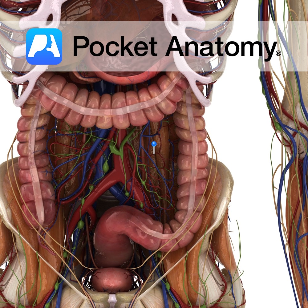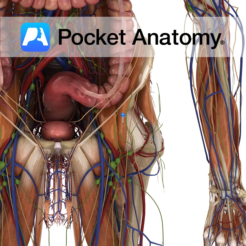
Anatomy
Origin:
Inner lip of Iliac crest, upper two-thirds of iliac fossa, ala of sacrum and anterior sacroiliac and iliolumbar ligaments.
Insertion:
Psoas tendon and lesser trochanter of femur.
Key Relations:
-Iliacus fills the iliac fossa of the hip bone and, enters the thigh with psoas major passing under the inguinal ligament.
-Forms the floor of the femoral triangle.
-The femoral nerve passes between the lateral border of the psoas major and iliacus muscles as it descends through the pelvis.
Note:
*Iliopsoas is a term which applies to the combined psoas major and iliacus muscles as they enter the thigh beneath the inguinal ligament and insert together onto the lesser trochanter.
Functions
-Flexes the thigh at the hip and stabilizes the hip joint.
-With psoas major medially and laterally rotates hip joint.
Supply
Nerve Supply:
Femoral Nerve (L2, L3).
Blood Supply:
-Iliac branches of iliolumbar artery
-Medial femoral circumflex artery.
Clinical
In rare cases the passage of the femoral nerve anterior to the iliacus becomes significant. The presence of a mass in the iliacus, such as a haematoma can cause compression of the femoral nerve. This leads to weakness of the quadriceps muscles of the anterior thigh, and loss of sensation in large areas of the anterior thigh and leg. Treatment to decompress the haematoma and remove the pressure on the femoral nerve can resolve the problem.
Interested in taking our award-winning Pocket Anatomy app for a test drive?
![]()
.jpg)
.jpg)