Anatomy
Origin:
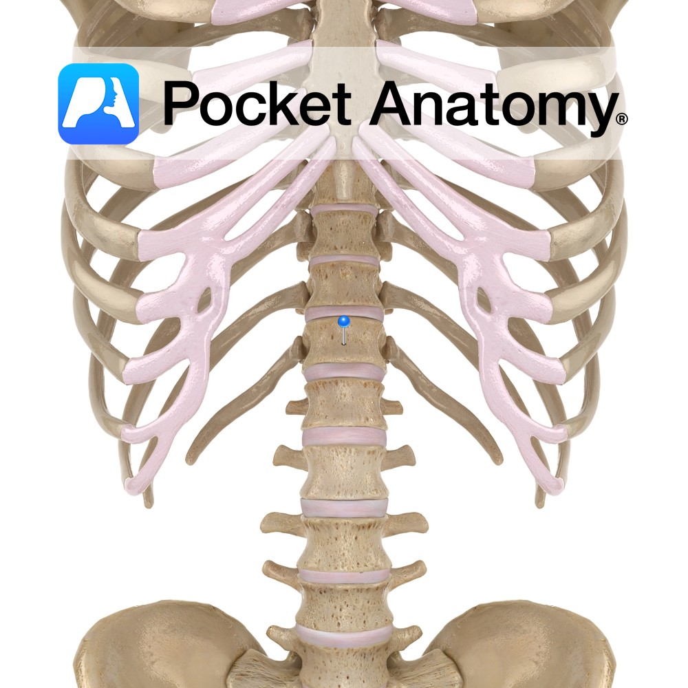
Sides of vertebra T12 to L5 and transverse processes of L1 to L5.
Insertion:
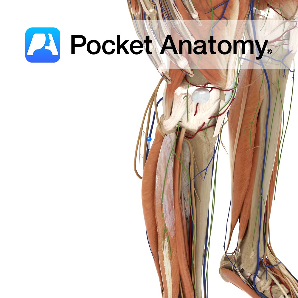
Lesser trochanter of the femur.
Key Relations:
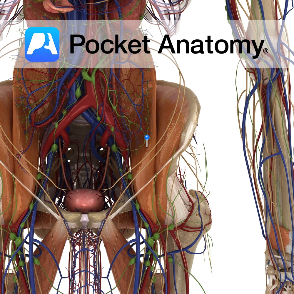
-The ureter passes along the medial aspect of the psoas major muscle as it enters the pelvis.
-Forms the floor of the femoral triangle.
-The femoral nerve (L2 to L4) travels through the substance of the psoas, before emerging to travel between the lateral border of psoas and the anterior surface of iliacus.
-The genitofemoral nerve (L1, L2) from the lumbar plexus emerges onto and runs down the surface of the psoas major muscle.
Note:
*Iliopsoas is a term which applies to the combined psoas major and iliacus muscles as they enter the thigh beneath the inguinal ligament and insert together onto the lesser trochanter.
Functions
-Flexes the thigh at the hip and stabilises the hip joint.
-With iliacus medially and laterally rotates hip joint.
-Important muscle in posture when standing.
Supply
Nerve Supply:
Ventral rami of lumbar spinal nerves 1, 2, 3.
Blood Supply:
Lumbar branches of the iliolumbar artery.
Clinical
Acute appendicitis is an abdominal emergency, caused by inflammation of the appendix. While the attachment of the appendix to the caecum is constant, the location of the remainder of the appendix varies between individuals.
The psoas sign is a finding on physical examination which can facilitate the diagnosis of appendicitis and indicates the appendix is located in a retrocaecal position. With a positive psoas sign the patient will feel increased abdominal pain when attempting to flex their right hip against resistance. This resistance is supplied by the physician’s hand which is placed just above the patient’s right knee. A retrocaecally located appendix may lie adjacent to the surface of the psoas muscle. An inflamed appendix in this position can irritate the muscle. This is the basis of the psoas sign.
Interested in taking our award-winning Pocket Anatomy app for a test drive?
![]()
.jpg)