Anatomy
Origin:
Spinous processes of T7 to T12 and the thoracolumbar fascia, by which it is attached to the spines of all lumbar and sacral vertebrae. The posterior part of the iliac crest and the lower three to four ribs.
Insertion:
The fibres converge to form a flattened tendon and attaches to the floor of the intertubercular sulcus of the humerus.
Key Relations:
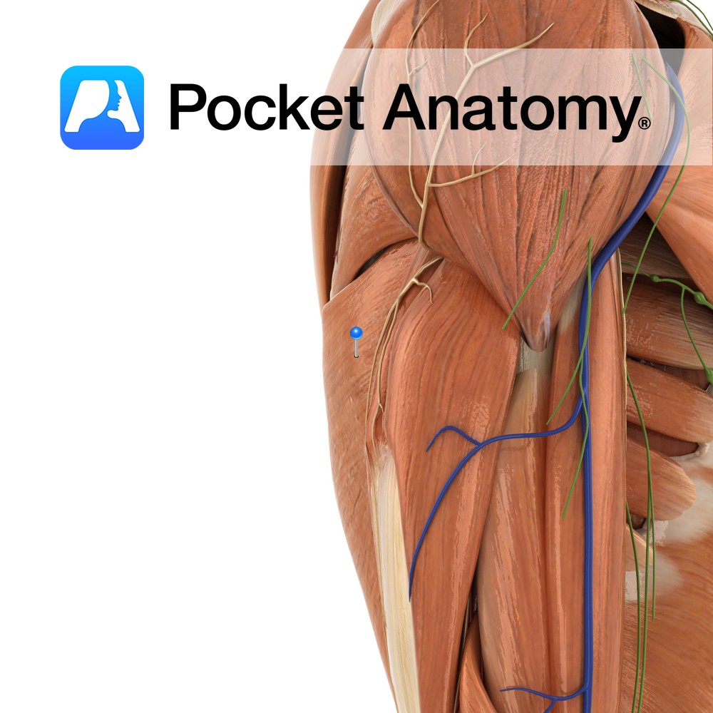
– The muscle curves around the inferolateral border of teres major to its anterior border.
-Along with teres major, it forms the posterior axillary fold.
-Is inferior border of the triangle of auscultation.
-Known as “The Lady Between Two Majors” due to its insertion between teres major and pectoralis major.
Functions
-Adducts, extends e.g. climbingand medially rotates the humerus at the shoulder joint.
-Draws shoulders down and back e.g. pushing on the arms of a chair when standing up.
-It is also an accessory muscle of respiration active in violent expiration.
Supply
Nerve Supply:
Thoracodorsal nerve (C6, C7, C8).
Blood Supply:
Thoracodorsal branch of the subscapular artery.
Clinical
It is the muscle most commonly used in cardiomyoplasty in which healthy muscle from another part of the body is wrapped around a failing heart along with a pacemaker to restore normal function.
Latissimus dorsi can be tested clinically provided the patient can start at a position whereby the arms are abducted above the head. They should then try and adduct the arms back down towards their torso against resistance from the examiner.
Interested in taking our award-winning Pocket Anatomy app for a test drive?
![]()