Anatomy
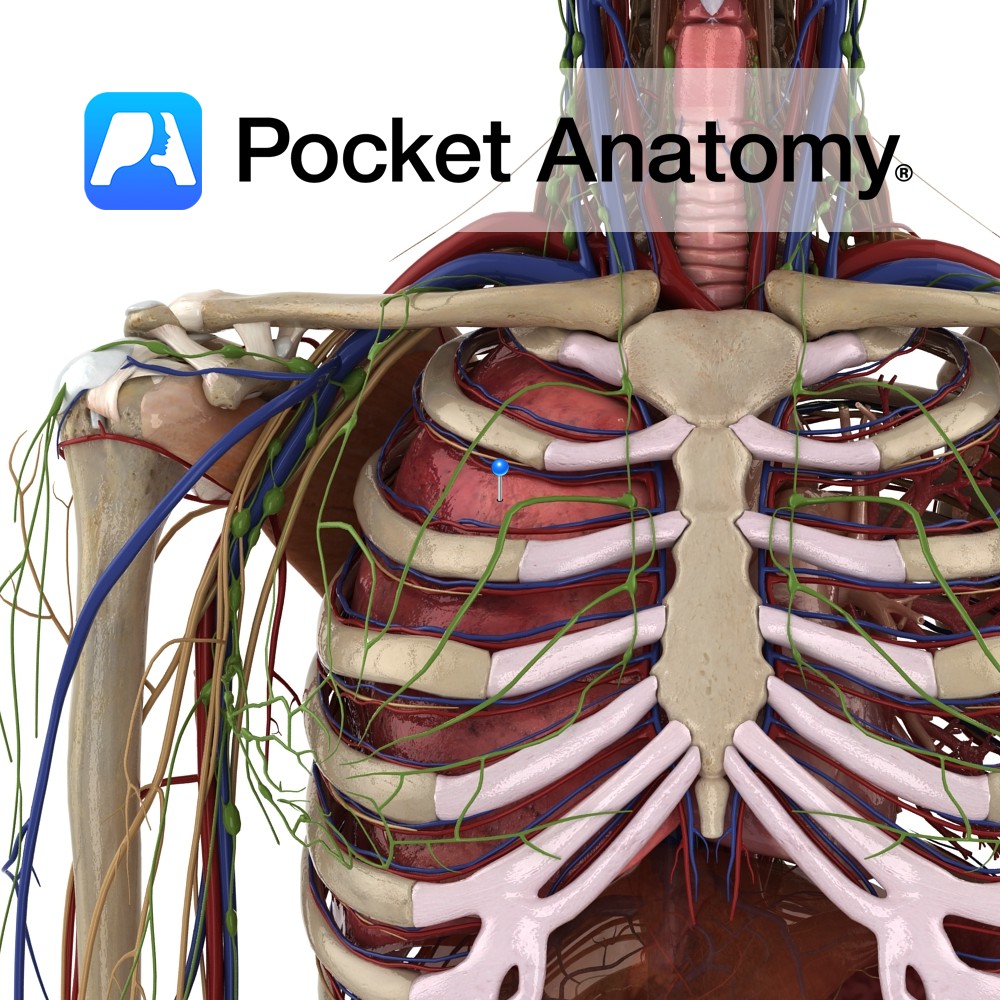
Paired thoracic respiratory organs, right 3 lobes, left 2 (heart accommodated), in closed pleural cavities, separated by mediastinum, diaphragm below, thoracic inlet (T1, 1st ribs, manubrium) above, surrounded by thoracic cage (T1-12, ribs, costal cartilages, sternum).
Hilum; artery, vein, bronchus. Trachea divides left and right bronchi, continues in lung: main bronchus – lobar (3R, 2L) – tertiary (supplying segments, 10 right, 8 left) – primary bronchiole – terminal – respiratory – alveolar ducts (gas exchange starts) – alveolar sacs – individual alveoli.
As bronchi branch, get smaller; more smooth muscle, less hyaline cartilage (rings proximally, isolated islands/plates distally), lining changes, ciliated mucous to squamous.
Clinical
On inspiration; diaphragm flattens itself (ie becomes less dome-like, effecting 75% of quiet breathing) thereby lengthening thoracic cavity, also intercostals (external, and interchondral part of internal) pull ribs out/up and increase cavity front-to-back (sternum-to-spine); parietal pleura attached to back of thoracic wall pulls pulmonary pleura by vacuum suction (lubricating serous allows pleura-on-pleura sliding); pulmonary pleura (attached to lung) thus expands lung; pressure drop inside sucks in air (conditioned progressively from nose/mouth); gaseous exchange happens – O2 in, CO2 out.
Expiration partly due to elastic recoil lung and pleura as diaphragm relaxes upwards, and abdominal muscles acting antagonistically to diaphragm.
Vignette
Air movement compromised: asthma (episodes of constriction bronchi/oles, different triggers); Chronic Obstructive Pulmonary Disease (COPD – emphysema and chronic bronchitis); bronchial/sac damage and scarring, difficulty exhaling); pulmonary fibrosis (stiff lungs, damage to sacs/interstitium). Infection: pneumonia (cough, chest pain, fever, dyspnea, streptococcus pneumoniae commonest agent), TB, ‘flu, fungal (in immunosuppression). Heart failure; heart can’t keep up with blood from lungs, backs up.
Cystic fibrosis, commonest in caucasians – 1 in 25 prevalence of autosomal recessive gene; thick mucus, recurrent infection, incremental damage. Cancer; 90% in smokers (30 times more than non-smokers), commonest site of metastases.
Interested in taking our award-winning Pocket Anatomy app for a test drive?
![]()