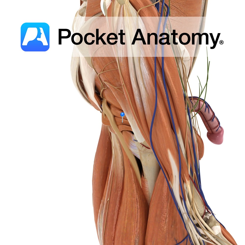
Anatomy
Origin:
Anterolateral wall of the pelvis, pelvic surface of the obturator membrane and margins of obturator foramen(i.e. inferior rami of pubis and ischium).
Insertion:
Medial surface of the greater trochanter of the femur.
Key Relations:
-Within the pelvis, forms lateral wall of ischiorectal fossa.
-Outside the pelvis, it’s tendon lies between the superior and inferior gemelli.
-The obturator canal is a passageway formed in the obturator foramen by part of the obturator membrane. It connects the pelvis to the thigh. The obturator artery, vein and nerve all pass through the canal.
Functions
-Laterally rotates the extended thigh at the hip joint.
-Abducts the flexed thigh at the hip joint.
Supply
Nerve Supply:
Nerve to obturator internus (L5, S1).
Blood Supply:
-Branches of the internal iliac arteries including the superior and inferior gluteal arteries
-Internal pudenal artery.
Interested in taking our award-winning Pocket Anatomy app for a test drive?
![]()