Anatomy
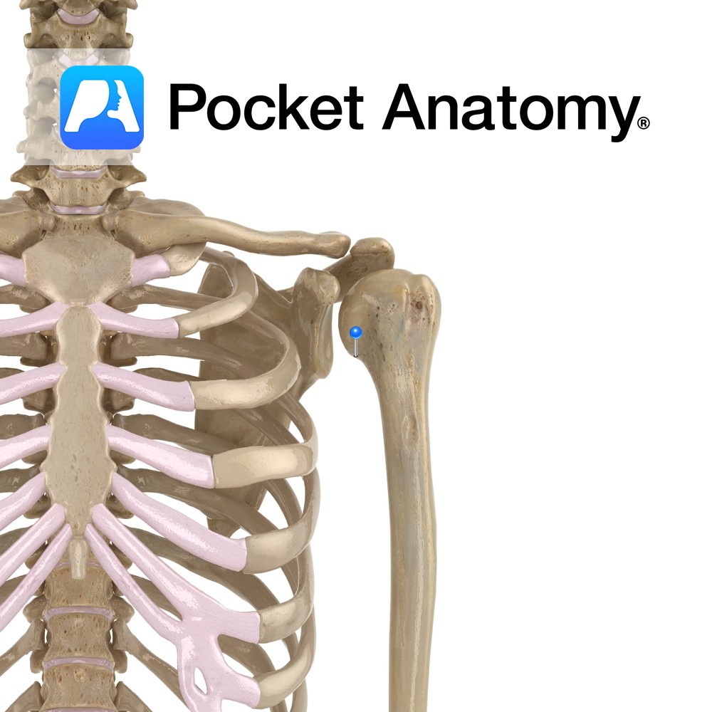
Origin:
Inferior aspect of the lateral supracondylar line of the femur and the popliteal surface of the femur.
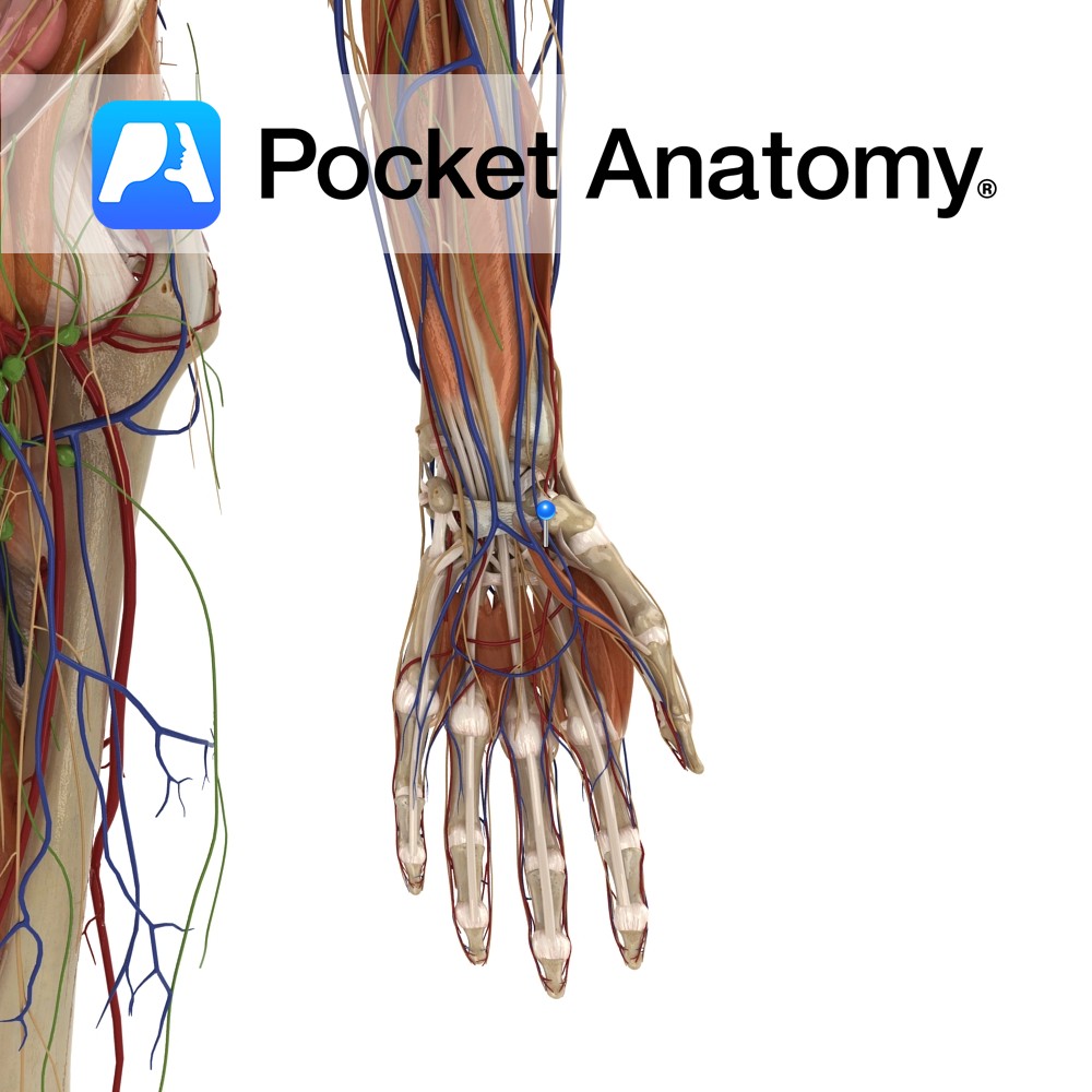
Insertion:
Medial side of posterior surface of calcaneus by the tendo calcaneus (Achilles tendon).
Key Relations:
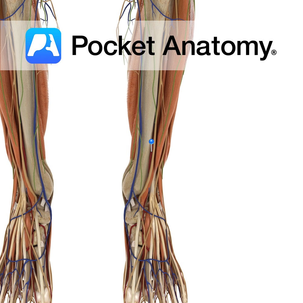
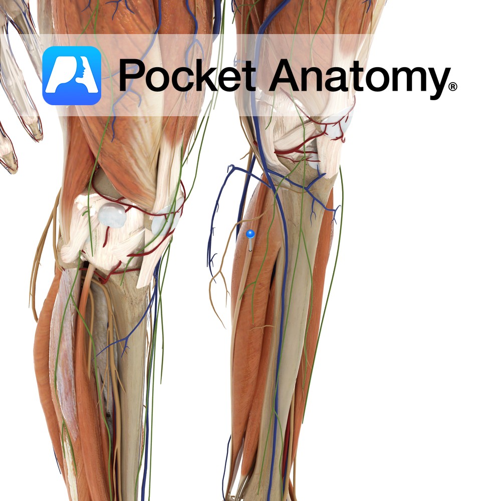
-One of the three muscles of the superficial posterior compartment of the leg.
-Forms the lateral lower border of the popliteal fossa.
Functions
-Weakly plantarflexes the ankle.
-Weakly flexes the knee joint.
(i.e. weakly assists gastrocnemius).
Supply
Nerve Supply:
Tibial nerve (S1, S2).
Blood Supply:
-Sural arteries from the popliteal artery
–Posterior tibial artery
-Fibular (peroneal) artery.
Clinical
The functional importance of plantaris is minimal therefore it is useful as a free tendon graft for reconstruction or reinforcement elsewhere, e.g. tendo calcaneus (Achilles tendon) repairs, with little functional deficit.
Interested in taking our award-winning Pocket Anatomy app for a test drive?
![]()