Anatomy
Course
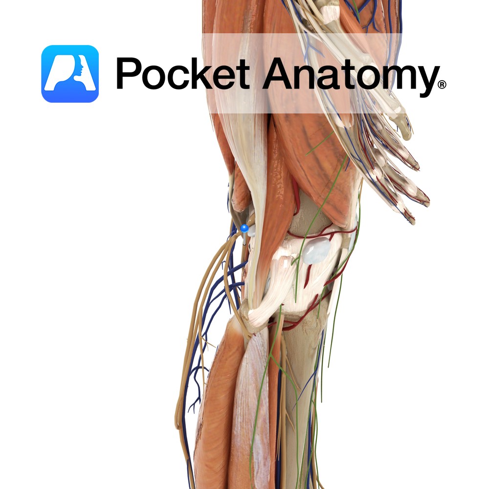
A terminal branch of the sciatic nerve, and so contains fibres from the spinal segments of L4 – S3. The sciatic nerve becomes the tibial nerve upon exiting the popliteal fossa.
It passes under the tendinous arch formed by the soleus muscle and descends in the plane between the solus and the tibialis posterior muscle. It enters the foot by passing behind the medial malleolus, through the tarsal tunnel and beneath the flexor retinaculum.
Supply
The tibial nerve supplies motor innervation to the muscles in the posterior compartment of the leg, as well as some sensory innervation via its branches; the sural and medial calcaneal nerve.
Clinical
Tarsal tunnel syndrome can arise as a result of damage to the tibial nerve, often caused by its compression when passing through the tarsal tunnel.
The symptoms include tingling, numbness or pain in the region of distribution of the tibial nerve, or a weakness in the muscles of the foot. It can be treated using pain management, or physical therapy, and in some cases surgery is required to widen the tarsal tunnel.
Some cases of tarsal tunnel resolve themselves without treatment.
Interested in taking our award-winning Pocket Anatomy app for a test drive?


