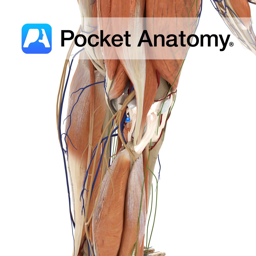
Anatomy
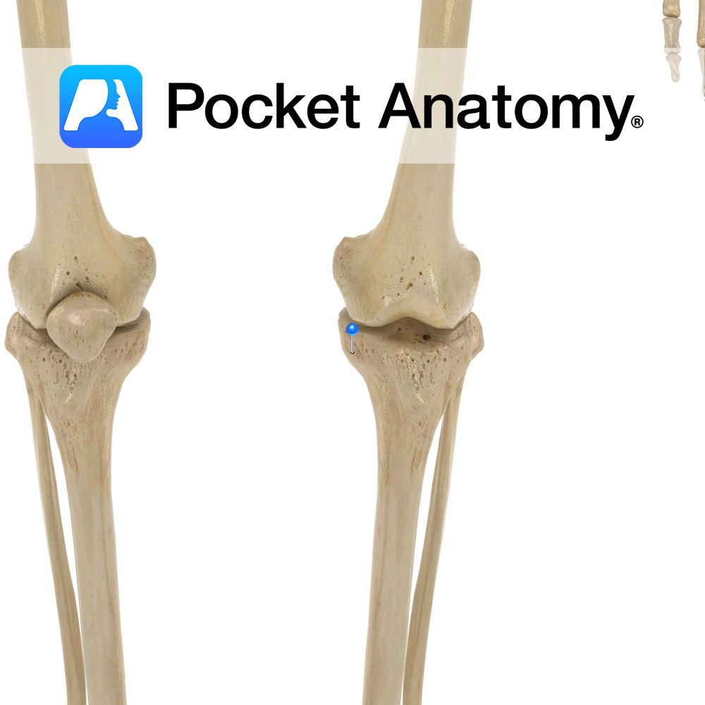
Origin:
Lateral surface of the lateral condyle of the femur and the lateral meniscus.
Insertion:
Medial half of the triangular area above the soleal line on the posterior surface of the tibia.
Key Relations:
-One of the four muscles of the deep posterior compartment of the leg.
-The popliteus forms part of the floor of the popliteal fossa.
-The popliteal tendon is intracapsular i.e it penetrates the capsule of the knee joint.
Functions
-Unlocks the extended knee by rotating the femur laterally on the fixed tibia.
-Weakly flexes the knee joint.
-Also stabilises the knee joint by aiding the posterior cruciate ligament to prevent forward dislocation of the femur.
Supply
Nerve Supply:
Tibial nerve (L4, L5, S1).
Blood Supply:
Popliteal artery.
Clinical
The popliteus muscle can be injured in a number of ways such as overuse, acute injury e.g a fall which may result in a rupture, surgery or swelling of the joint which may increase tone (tightening) of the muscle or weakened quadriceps or hamstrings which may also increase tone. Rupture of the popliteus muscle often occurs in conjunction with rupture of the posterior cruciate ligament. The patient may present with posterior knee pain, pain on palpation and/or pain on resisted flexion of the knee joint.
Treatment includes rest, NSAIDs, stretching of the hamstrings and a physiotherapy programme to strengthen the quadriceps and hamstrings.
Interested in taking our award-winning Pocket Anatomy app for a test drive?
![]()
.jpg)
.jpg)