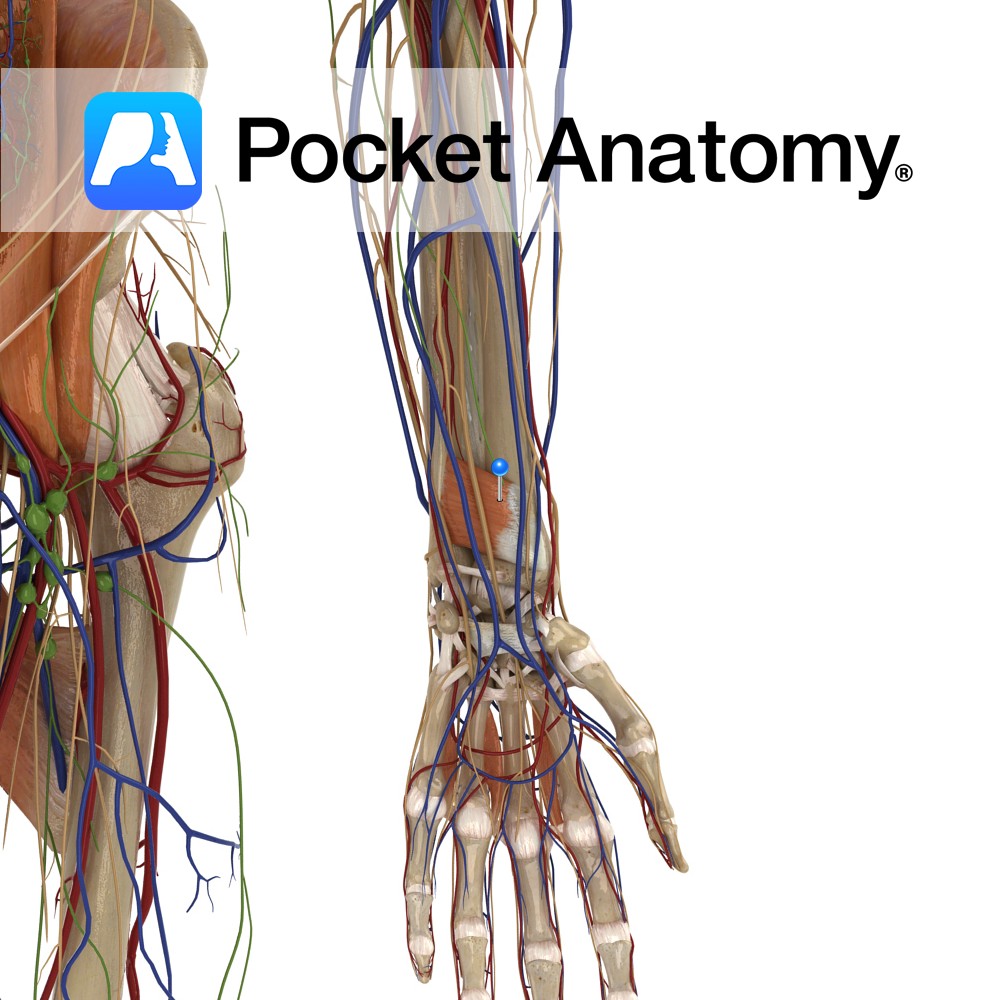
Anatomy
Origin:
Medial surface of distal ulna.
Insertion:
Lateral Surface of distal radius.
Key Relations:
One of the three muscles in the deep anterior compartment of the forearm.
Functions
-Pronates the forearm and hand at the radioulnar joint.
-Deep fibres bind the radius and ulna together.
e.g.: as in loosening a screw (right hand).
Supply
Nerve Supply:
Anterior Interosseus nerve (C8,T1), branch of Median nerve.
Arterial Supply:
Anterior Interosseous artery.
Clinical
Pronator quadratus has a well defined fascial covering. This creates a space in which fluid can accumulate. On most normal radiographs of the distal lateral forearm a thin radiolucency is seen representing a layer of fat overlying the fascial covering of pronator quadratus. If there is an accumulation of fluid within the fascial compartment of the muscle the layer of fat can be displaced. When observed on a radiograph the displaced radiolucency is known as the pronator quadratus sign.
Interested in taking our award-winning Pocket Anatomy app for a test drive?
![]()