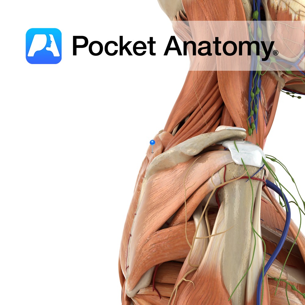
Anatomy
Origin:
Lower ligamentum nuchae and the spinous processes of C7 to T1.
Insertion:
Medial border of the scapula at the level of the spine, superior to the attachment of rhomboid major.
Key Relations:
-Superior to rhomboid major.
-Posterior to trapezius.
-Fascia of rhomboid minor and serratus anterior are tightly fused.
Functions
Retracts e.g. pulling open a drawer and stabilizes the scapula to the thoracic wall.
Supply
Nerve Supply:
Dorsal scapular nerve (C4, C5).
Blood Supply:
-Dorsal scapular artery
-Dorsal perforating branches of the upper 5-6 posterior intercostal arteries.
Clinical
As the scapula does not have a bony attachment to the torso, it is stabilized by muscles such as rhomboid minor attached between its medial border and the spine. Damage to rhomboid minor can therefore cause scapular instability.
Rhomboid minor can be tested clinically by gross observation. If function is decreased, the scapula on the affected side may lie more lateral than the scapula of the fully.
Interested in taking our award-winning Pocket Anatomy app for a test drive?
![]()