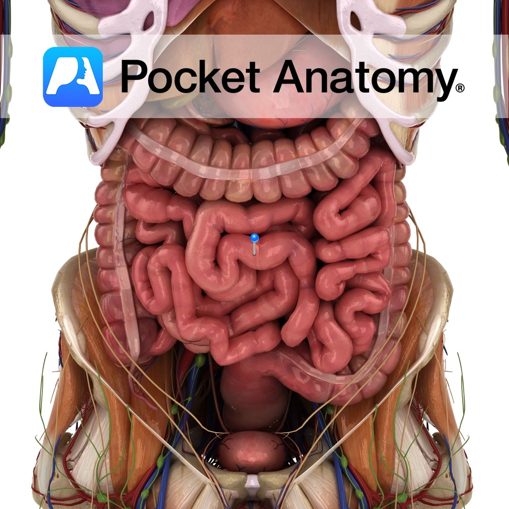
Anatomy
Between stomach and colon (pyloric sphincter to ileocecal valve), comprising duodenum, jejunum and ileum, convoluted coils occupying much of abdomen/pelvis, framed at sides and above by colon, c. 23′ long in adult), attached to posterior abdominal wall by fan-shaped mesentery (invaginated peritoneum, containing lymph nodes and vessels) other than duodenum (mostly retroperitoneal).
Supplied by superior mesenteric artery (from aorta, behind neck pancreas) – anastomosing jejunal/ileal branches – vasa recta (end branches). Veins to superior mesenteric vein – with splenic vein (joined by inferior mesenteric vein) to form portal vein – liver. Jejunal coils central, left upper abdomen; ileal central, right lower abdomen and pelvis.
Clinical
SI longer than LI, called “small” as lumen smaller – little more than 1″. Digestion is breakdown of food into components then absorbed; mouth – mastication, saliva, bolus on through esophagus; stomach – gastric juice with pepsin, HCl, 1-2 hours, on through pylorus; duodenum – pancreatic enzymes; jejunum – further digestion, absorption; ileum – by end, 95% absorption complete).
Interested in taking our award-winning Pocket Anatomy app for a test drive?
![]()
.jpg)