Anatomy
Origin:
Posterior surfaces of the head and upper third of the fibula, the soleal line of the tibia and the middle third of the medial border of the tibia.
Insertion:
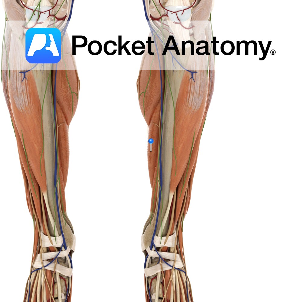
Middle part of the posterior surface of the calcaneus by the tendo calcaneus (Achilles tendon).
Key Relations:
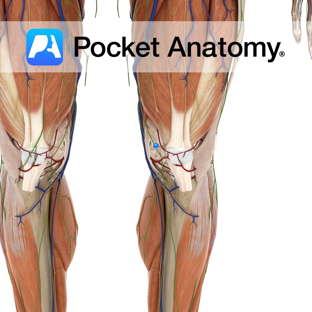
-One of the three muscles of the superficial posterior compartment of the leg.
-The soleus gives rise to a tendon called the tendo calcaneus (Achilles tendon) which fuses with the gastrocnemius.
-The two heads of gastrocnemius along with soleus forms a muscular mass also known as the ‘triceps surae’ which makes up the majority of the calf.
Functions
-Plantarflexes the ankle joint with gastrocnemius through the tendo calcaneus (Achilles tendon) e.g. standing on tip toes.
-Prevents excessive dorsiflexion during walking.
-Stabilises the leg over the foot e.g. standing upright..
Supply
Nerve Supply:
Tibial nerve (S1, S2).
Blood Supply:
-Sural arteries from the popliteal artery
–Posterior tibial artery
-Fibular (peroneal) artery.
Clinical
Calf strain or rupture is a common sports injury which can affect either of the two calf muscles- gastrocnemius or soleus. Calf strain or rupture most commonly occurs in racquet sports, basketball, running or skiing. The patient often presents with a sharp sudden calf pain, tightness followed by swelling and ecchymosis. It is graded according to the severity of the symptoms with grade 1 being a minor rupture and grade 3 being a 90% or full rupture. Treatment includes RICE (rest, ice, compression and elevation), NSAIDs and sports massage or physiotherapy programme.
Interested in taking our award-winning Pocket Anatomy app for a test drive?
![]()