Anatomy
Origin:
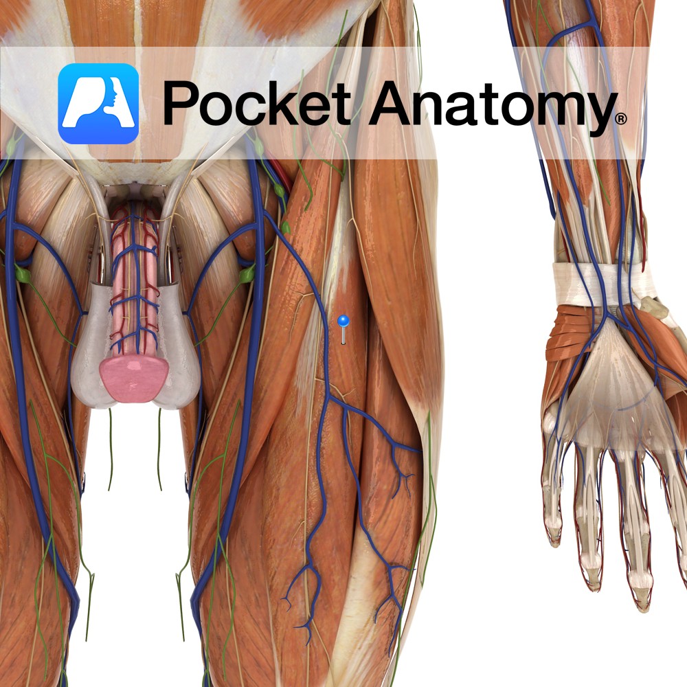
Straight head: anterior inferior iliac spine.
Reflected head: a groove on the upper surface of the acetabulum and from the fibrous capsule of the hip joint.
Insertion:
Base of the patella via the quadriceps femoris tendon. The quadriceps femoris tendon is functionally continuous with the patellar ligament which runs from the apex of the patella to the tibial tuberosity.
Key Relations:
-One of the five muscles of the anterior compartment of the thigh.
-From its separate origins the 2 heads combine to form a single muscle belly lying anterior to vastus intermedius, and between the vastus lateralis and vastus medialis muscles.
Note:
The quadriceps femoris is a large muscle group containing the 3 vastus muscles and the rectus femoris muscle of the anterior thigh. The quadriceps femoris tendon represents their shared insertion onto the patella.
Functions
-Together with the other muscles that make up quadriceps femoris extends the leg at the knee joint.
-Flexes the thigh at the hip joint.
Supply
Nerve Supply:
Femoral nerve (L2, L3, L4).
Blood Supply:
-Lateral circumflex femoral artery
-Deep femoral artery.
Clinical
Testing the patellar reflex: a tap of a tendon hammer on the patellar ligament should elicit a brief contraction of the quadriceps femoris muscles. This is a reflex action in response to the stretching of muscle spindles within the muscles. The spinal segments L3 and L4 predominantly innervate the quadriceps femoris muscles, and testing of the patellar reflex assesses reflex activity at these levels of the spinal cord.
Interested in taking our award-winning Pocket Anatomy app for a test drive?


