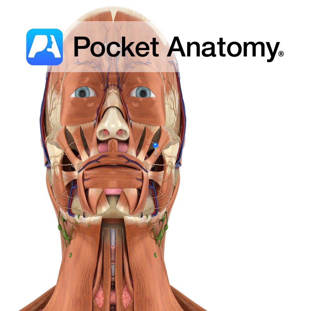
Anatomy
Origin:
Posterior part of the lateral surface of the zygomatic bone.
Insertion:
Angle of mouth where its fibres blend with those of levator anguli oris, depressor anguli oris and orbicularis oris.
Key Relations:
Arises posterior to orbicularis oculi and runs downwards and blends with orbicularis oris before inserting on the angle of the mouth.
Functions
Draws the angle of the mouth upwards and backwards e.g. as in laughing or smiling..
Supply
Nerve Supply:
Buccal branch of the facial nerve (CN 7).
Blood Supply:
Superior labial branch of facial artery.
Clinical
A common variation in the population is a ‘double’ or ‘bifid’ zygomaticus major muscle. In this case the muscle divides into two bundles midway from its origin to its insertion. In some cases this inferior bundle has a dermal attachment midway along its length. This particular variation is thought to be responsible for the formation of cheek dimples.
Interested in taking our award-winning Pocket Anatomy app for a test drive?
![]()
.jpg)