
PocketAnatomy® is a registered brand name owned by © eMedia Interactive Ltd, 2009-2027.
iPhone, iPad, iPad Pro and Mac are trademarks of Apple Inc., registered in the U.S. and other countries. App Store is a service mark of Apple Inc.
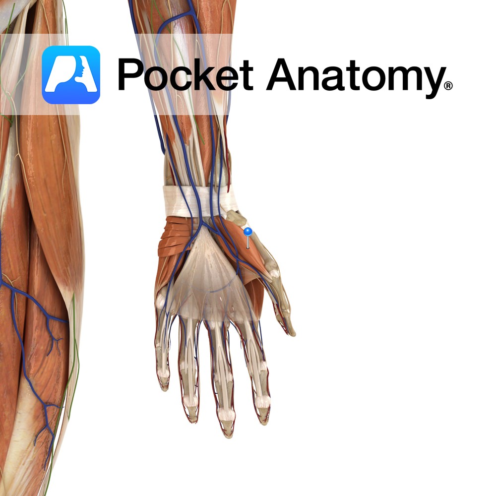
Anatomy Origin: Tubercles of scaphoid and trapezium and adjacent flexor retinaculum. Insertion: Proximal phalanx and extensor apparatus of the thumb. Key Relations: Is one of the muscles of the thenar eminence of the hand. Functions -Abduction of the thumb at the carpopmetacarpol and metacarpophalangeal joints. -Also assists in thumb opposition and extension. Supply Nerve Supply:
- Published in Pocket Anatomy Pins
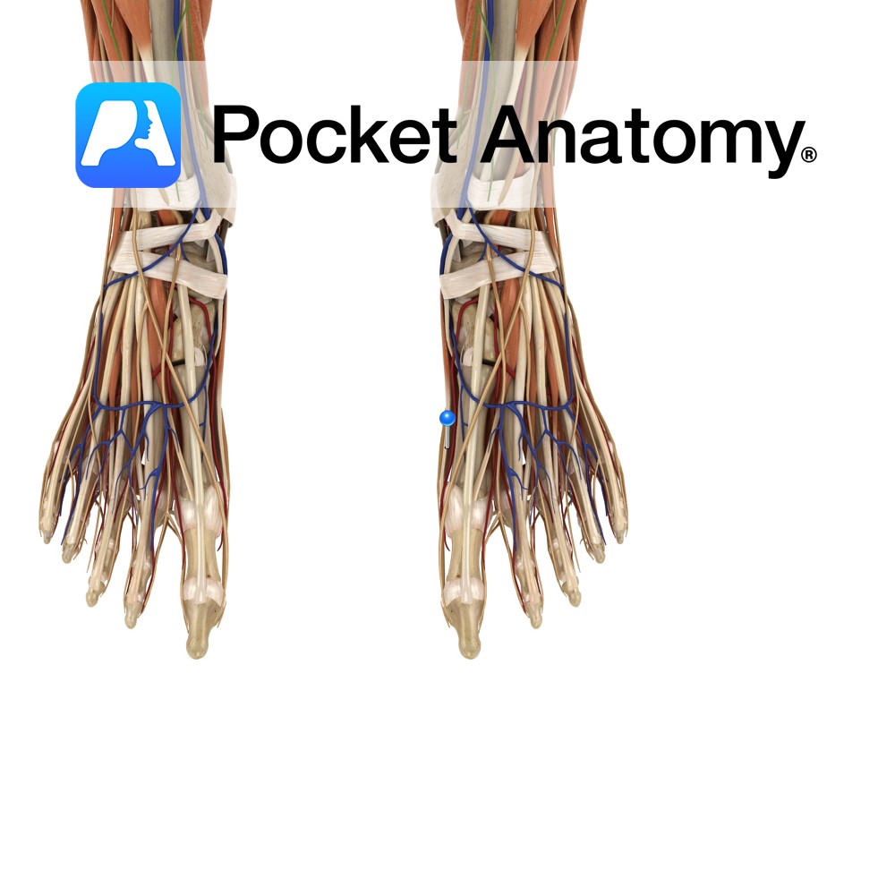
Anatomy Origin: Medial process of the calcaneal tuberosity, flexor retinaculum and plantar aponeurosis. Insertion: Medial surface of the base of the proximal phalanx of the hallux (big toe). Key relations: Lies on the medial surface of the foot and contributes to a soft tissue bulge on the medial part of the sole of the foot.
- Published in Pocket Anatomy Pins

Anatomy Origin: Pisiform and tendon of flexor carpi ulnaris. Insertion: Ulnar aspect of the base of the proximal phalanx of the little finger and ulnar border of the extensor apparatus of the little finger. Key Relations: Is one of the muscles of the hypothenar eminence of the hand. Functions Abduction of the little finger at
- Published in Pocket Anatomy Pins

Anatomy Origin: Lateral and medial processes of the calcaneal tuberosity and plantar aponeurosis. Insertion: Lateral surface of the base of the 5th proximal phalanx. Key relations: -Lies on the lateral surface of the foot. -The lateral plantar vessels and nerve located medially. Functions Abducts the 5th toe at the metatarsophalangeal joint. Supply Nerve supply: Lateral
- Published in Pocket Anatomy Pins
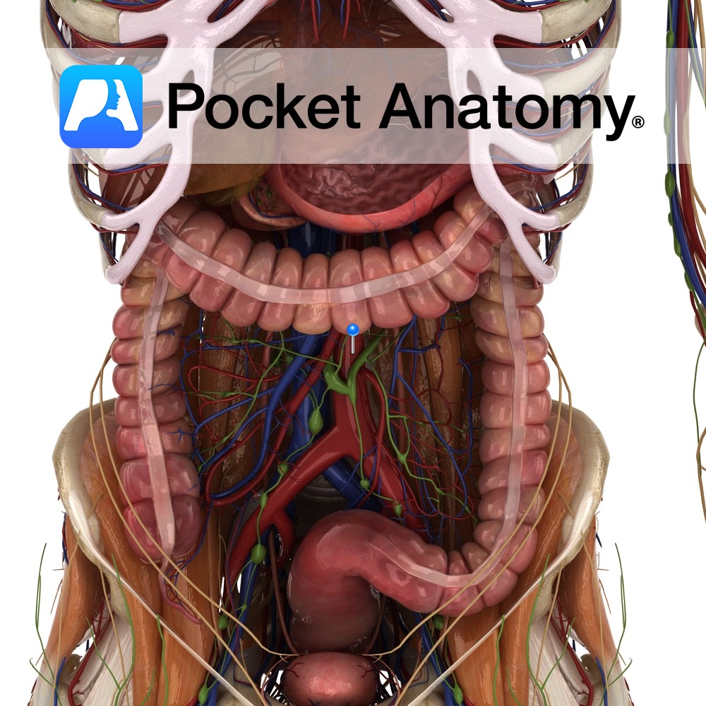
Anatomy Course Enters the abdomen as a continuation of the thoracic aorta by passing through the aortic hiatus of the diaphragm at the level of T12. Descends downward slightly to the left of the midline until it bifurcates into the left and right common iliac arteries at the level to L4. The inferior vena cava
- Published in Pocket Anatomy Pins
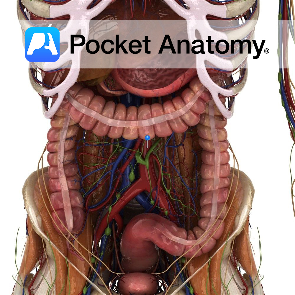
Course Enters the abdomen as a continuation of the thoracic aorta by passing through the aortic hiatus of the diaphragm at the level of T12. Descends downward slightly to the left of the midline until it bifurcates into the left and right common iliac arteries at the level to L4. The inferior vena cava travels
- Published in Pocket Anatomy Pins
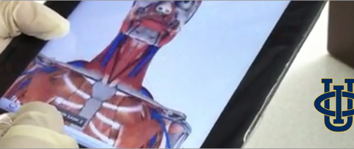
“…there are quite a few apps that effectively marry anatomy content and the iPad technology such as the Pocket Anatomy app.” Dr. Warren Weichmann, University of California Irvine.
- Published in Main Blog, Teaching Anatomy

Hello! Pocket Anatomy is there on your iPad and you get excited every time you show it to your friends. The coolest part? Flipping through the two human models showing the layers and the beautiful 3D graphic details. Our Interactive 3D Human Body app is designed in such a way to allow you to navigate and
- Published in Main Blog, Teaching Anatomy, Tutorial

Last month we started to see how medical terminology, no matter how complex it looks like, can be decoded by becoming more familiar with words roots deriving from -mostly – Greek and Latin. So far we focused only on parts of the human body. This month, we will look at more general concepts, like diseases
- Published in Main Blog, Teaching Anatomy, Tutorial
So, you’ve just downloaded the Pocket Anatomy app? You’re maybe spending hours spinning around the 3D models of the male and female body and have certainly been admiring the beauty of them. You’re amazed at how complex this machine is! Now, it’s high time to understand how the human body works and how each single part
- Published in Main Blog, Teaching Anatomy, Tutorial