PocketAnatomy® is a registered brand name owned by © eMedia Interactive Ltd, 2009-2022.
iPhone, iPad, iPad Pro and Mac are trademarks of Apple Inc., registered in the U.S. and other countries. App Store is a service mark of Apple Inc.
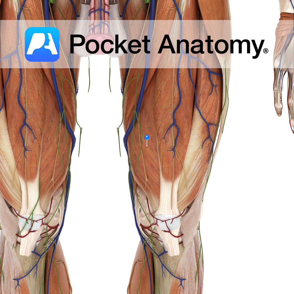
Anatomy Origin: Intertrochanteric line and medial lip of linea aspera of femur. Insertion: Base of the patella via the quadriceps femoris tendon. The quadriceps femoris tendon is functionally continuous with the patellar ligament which runs from the apex of the patella to the tibial tuberosity. Key Relations: -One of the five muscles of the anterior
- Published in Pocket Anatomy Pins
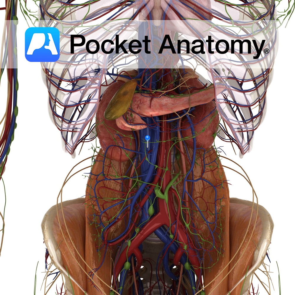
Anatomy Course The main vein of the body. It begins when the two common iliac veins unite at the approximate vertebral level of L5. It ascends on the right side of the vertebral column, retroperitoneally and on the posterior abdominal wall. It receives a number of tributaries along its way. It drains directly into the
- Published in Pocket Anatomy Pins
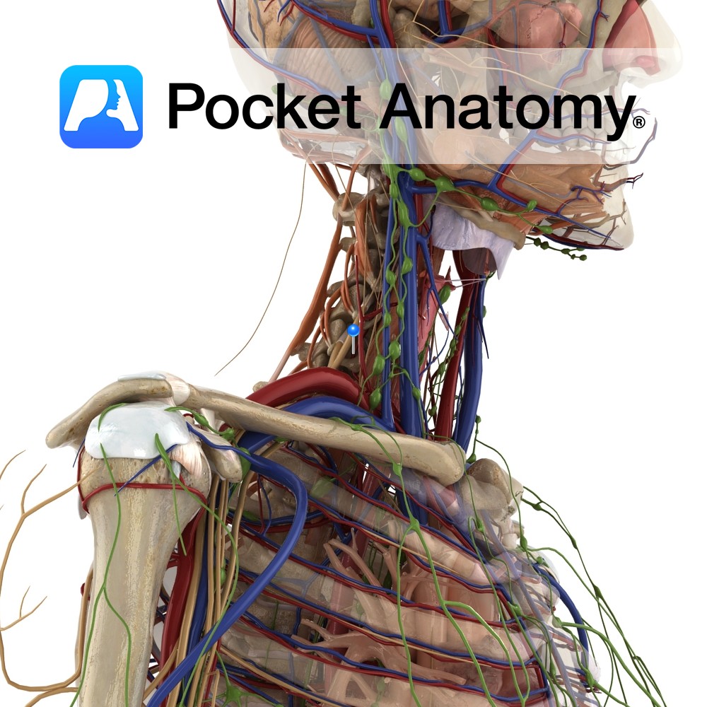
Anatomy Course Arise from the subclavian arteries and ascend through the neck, passing through the transverse foramina of the cervical vertebrae. They enter the cranial vault through the foramen magnum, and eventually unite to form the basilar artery at the base of the medulla. Supply Contribute to the blood supply of the brain. Interested in
- Published in Pocket Anatomy Pins
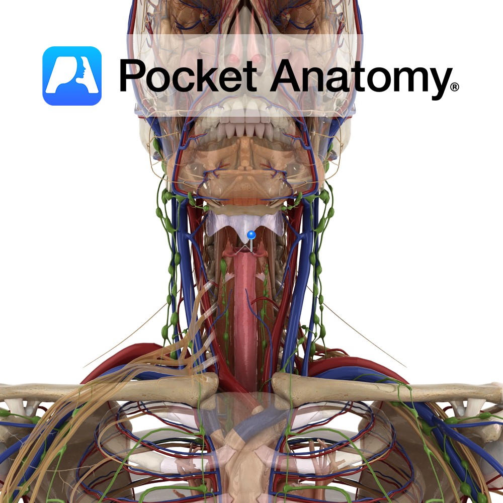
Anatomy The vocal ligaments are two bands of elastic tissue enclosed in the vocal folds. They attach to the posterior side of the thyroid cartilage anteriorly and the vocal process of the vocal process of the arytenoids cartilage. Functions Part of the larynx which is both a valve to close the lower respiratory and an
- Published in Pocket Anatomy Pins
Anatomy Muscular sheet across lower (outlet) pelvis; superficial transversus perinei, bulbospongiosus muscles (around base penis), ischiocavernosus muscle, sphincter urethrae (at membranous urethra); all variably involved in closing/opening by compression/relaxation. Interested in taking our award-winning Pocket Anatomy app for a test drive?
- Published in Pocket Anatomy Pins
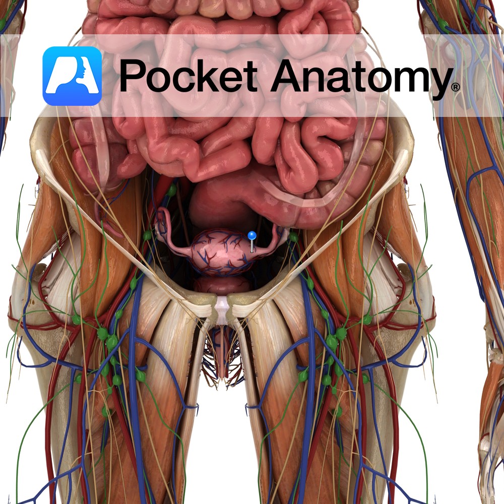
Anatomy The meeting of uterus (lined by endometrium) and fallopian tube (lined by ciliated epithelium). Physiology The part of the fallopian tube within the uterine wall – interstitial – is both narrow and muscular, and may play a role in impeding the upward progress of faulty sperm, and in delaying the downward progress of the
- Published in Pocket Anatomy Pins
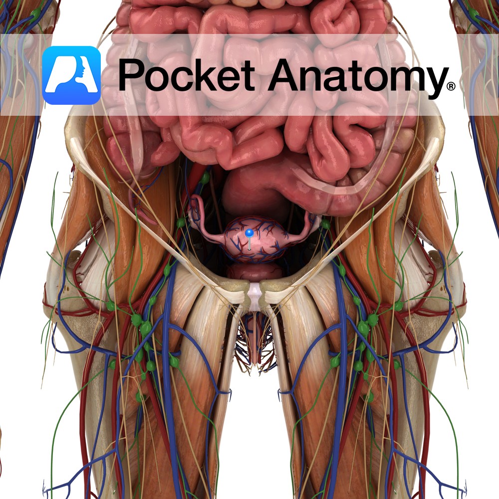
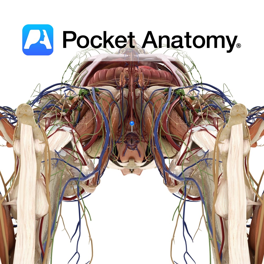
Anatomy Hollow thick-walled pear-shaped muscular pelvic (and also abdominal when gravid/pregnant) reproductive organ, approximately 7cms long X 5 wide when non-gravid/empty. Body (upper 2/3 of uterus/womb) rounded at top (fundus) where it is joined at the side/cornu/horn by fallopian tubes, uterine cavity narrows at bottom of body (isthmus) at beginning of cylindrical neck/cervix (lower 1/3)
- Published in Pocket Anatomy Pins
Anatomy Pelvic female sex organ, a fibromuscular tube (lower part of genital tract and birth canal), courses up and back, connects vulva (behind urethral orifice, protected by labia) to uterus (at external os of cervix), behind bladder (and urethra and distal ends ureters) and upper part of cervix, in front of rectum, between levators ani,
- Published in Pocket Anatomy Pins
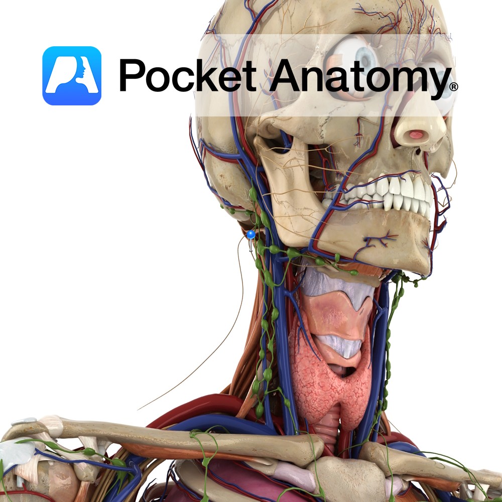
Anatomy Course The Vagus Nerve (X) is the tenth cranial nerve, and it originates from several nerve rootlets in the brain on the anterolateral surface of the medulla oblongata. These rootlets unite to form the vagus nerve before exiting the skull through the jugular foramen. It then descends through the neck within the carotid sheath.
- Published in Pocket Anatomy Pins
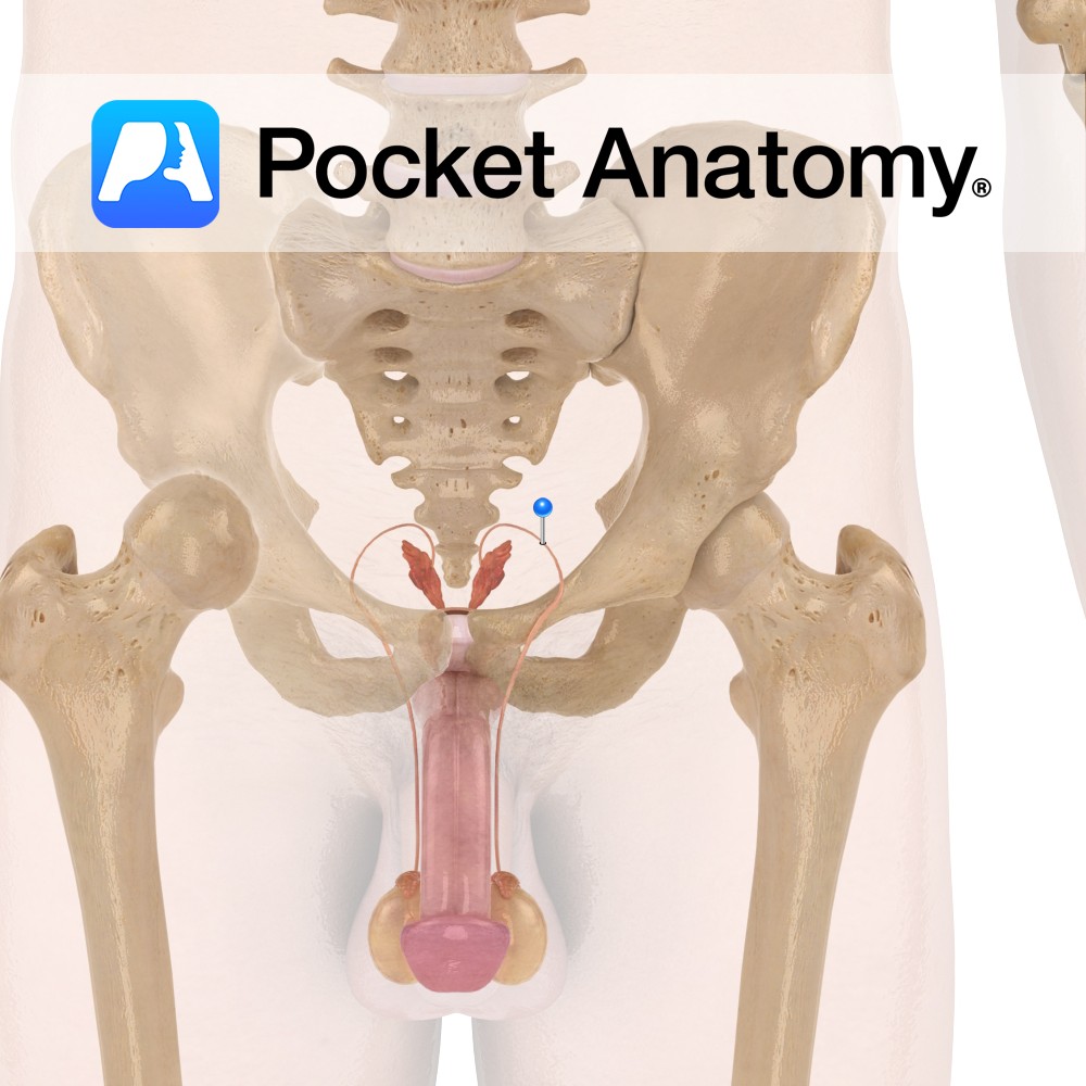
Functions Moves sperm from epididymis to ejaculatory ducts (by means of smooth muscle peristalsis) in anticipation of ejaculation. Anatomy Paired structure of male reproduction, connects the epididymis to ejaculatory ducts for the movement of sperm. Usually ~30cm long, 3-5mm diameter, contain smooth muscle. An ampulla (tortuous in shape) is often seen at the efferent end,
- Published in Pocket Anatomy Pins