Anatomy
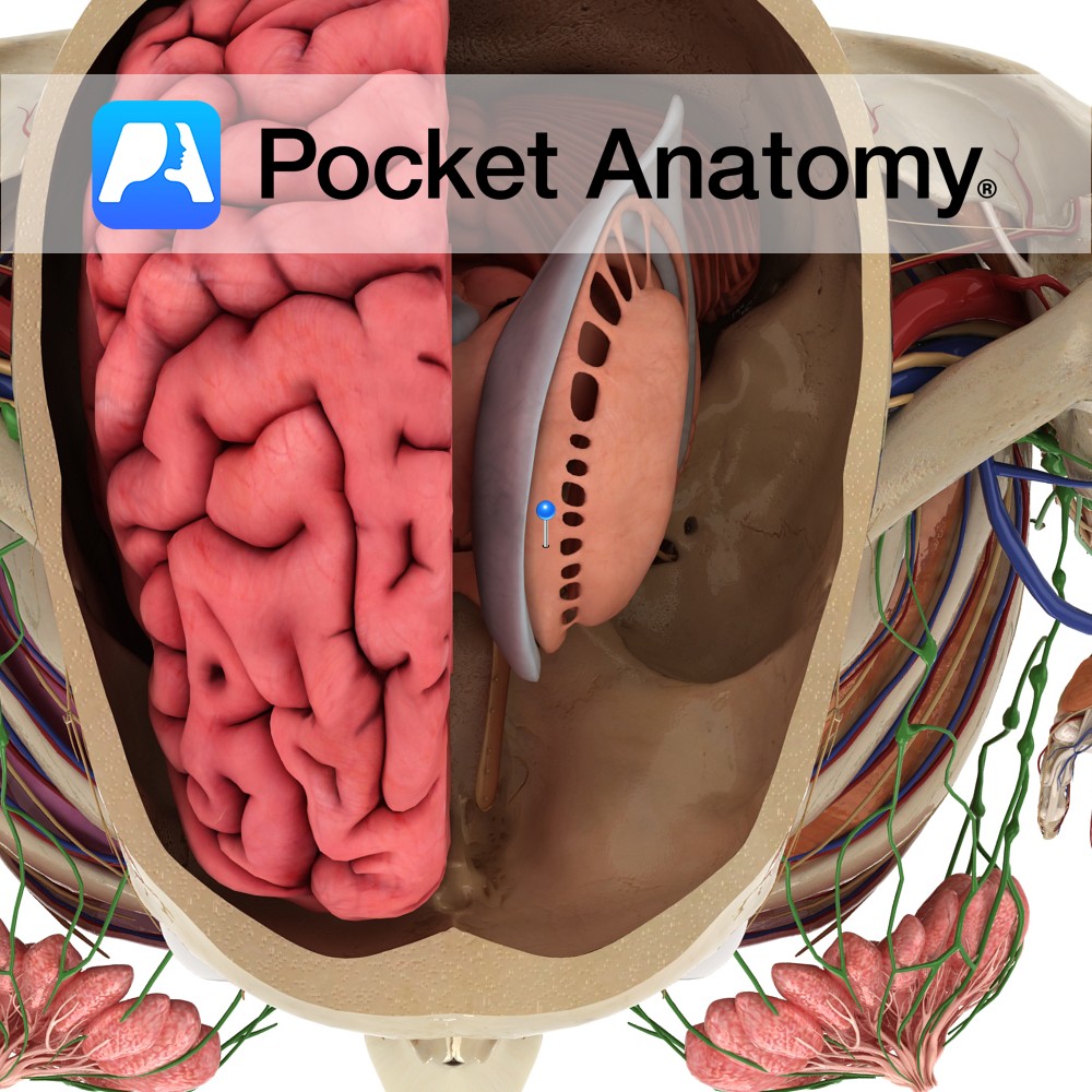
The caudate nucleus comprises a head, body and tail. The head of the caudate is rounded and forms the anterior portion of the wall of the lateral ventricule. Its fibers pass through the internal capsule to be in continuity with the putamen.
The body of the caudate is continuous with the head, adjacent to the interventricular foramen of Monro. It forms part of the floor of the lateral ventricle.
The tail of the caudate follows the contour of the lateral ventricle. It tapers gradually on descent into the temporal lobe and the amygdala marks its point of termination.
Blood Supply:
Supplied by branches of the internal carotid, anterior cerebral and middle cerebral arteries.
Functions
The caudate nucleus and putamen have common functions and are collectively known as the striatum. The striatum is the main receiver of information in the basal ganglia.
The caudate has also been implicated in the modulation of feedback between the subcortex and cortex, to ensure that fulminant activation does not occur. It is also thought to have functions that relate to learning and memory, and has been linked to regions of the frontal lobe that are responsible for executive functions.
Afferent pathways:
-Corticostriatal projection: Fibers originate extensively from the ipsilateral cerebral cortex. They terminate largely in the putamen and use the excitatory neurotransmitter glutamate.
-Thalamostriatal projection: Fibers originate from the intralaminar nuclei of the ipsilateral thalamus. They use the excitatory neurotransmitter glutamate.
-Nigrostriatal projection: Fibers originate from the pars compacta of the substantia nigra. They use dopamine as their neurotransmitter, which may be excitatory or inhibitory.
Efferent pathways:
-Striatopallidal projection: Fibers project to the globus pallidus and utilise the inhibitory neurotransmitter GABA.
-Striatonigral projection: Fibers project to the to the pars reticulata of the substantia nigra and utilise the inhibitory neurotransmitter GABA.
Both efferent pathways also use neuropeptides to modulate their responses e.g. substance P and dynorphin.
Clinical
Parkinson’s Disease (PD) arises from a deficiency of dopamine in the caudate and putamen, caused by the degeneration of dopaminergic neurons in the pars compacta of the substantia nigra. This damage reduces the activity of the Direct Pathway and increases the activity of the Indirect Pathway, ultimately impairing movement.
Huntington’s Disease (HD) arises from the accumulation of the mutant protein ‘Huntingtin’, causing degeneration in the caudate and putamen. This damage increases the activity of the Direct Pathway and decreases the activity of the Indirect Pathway, ultimately increasing movement. Huntington’s disease results in chorea, psychiatric changes, and progressive cognitive impairment.
An isolated caudate lesion may sometimes give rise to chorea.
Interested in taking our award-winning Pocket Anatomy app for a test drive?
![]()