Anatomy
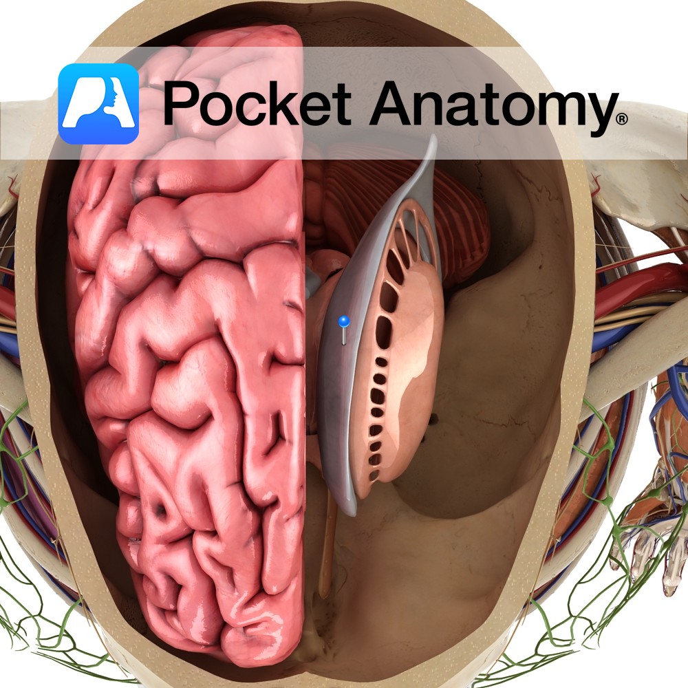
This is a C-shaped cavity within each cerebral hemisphere lined with ependymal cells. They are part of the series of fluid-filled cavities, which make up the ventricular system. It is connected to the third ventricle via the interventricular foramen of Monro.
It divides into a body, which occupies the parietal lobe and anterior, posterior and inferior horns, which extend into the frontal, occipital and temporal lobes respectively. The body extends from the interventricular foramen of Monro to the posterior end of the thalamus joining the anterior horn with the posterior and inferior horns.
The roof is formed by the corpus callosum. The floor is the body of the caudate nucleus and lateral margin of the thalamus. The medial wall is the septum pellucidum.
Functions
Filled with cerebrospinal fluid (CSF) and drains CSF to the third ventricle.
Clinical
Hydrocephalus denotes ventricular enlargement. Hydrocephalus may be communicating (i.e not obstructive) e.g. normal pressure hydrocephalus, or non-communicating (i.e. obstructive hydrocephalus) e.g. colloid cyst obstructing the third ventricle. Normal pressure hydrocephalus is associated with gait impairment, cognitive impairment, and urinary incontinence.
Hydrocephalus ex-vacuo may occur when the ventricles enlarge to fill some of the space created by cerebral atrophy.
Interested in taking our award-winning Pocket Anatomy app for a test drive?
![]()