Motion
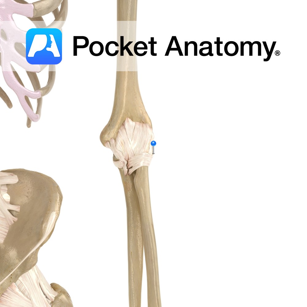
The elbow joint is a uniaxial compound synovial hinge joint. The elbow joint actually includes 2 joints the humeroulnar joint and the humeroradial joint. The capitulum of the humerus articulates with the head of the radius, and the trochlea of the humerus articulates with the trochlear notch of the ulna. Together these articulations make up the elbow joint. The elbow joint permits flexion and extension only. The trochlea has an asymmetrical structure, which creates angulation of the ulna when extended (i.e. the forearm deviates laterally from the arm) forming a ‘carrying angle’. This allows the forearms and hands to hang away from the hips when for example carrying bags.
* The proximal radioulnar joint is also contained within the synovial membrane of the elbow, but is dealt with separately here.
Stability
The fibrous capsule surrounding the joint plays a role in its stability, it is thinner posteriorly but is reinforced (see below)
The capsule is further strengthened by a number of muscles and tendons.
-Anteriorly it receives fibres from brachialis
-Posteriorly it is related to the tendons of triceps and anconeus.
-Laterally it is related to supinator and the common extensor tendon.
-Medially it is related to the common flexor tendon and flexor carpi ulnaris
The ligaments important in stabilizing the elbow joint include:
-Radial (lateral) collateral ligament
-Ulnar (medial) collateral ligament
–Annular ligament.
Muscles
Flexion:
Biceps brachii
Brachialis
Brachioradialis
Extension:
Triceps brachii
Anconeus.
Clinical
The most common injuries of the elbow are overuse injuries such as tennis elbow and golfer’s elbow. Tennis elbow is inflammation (tendinitis) of the common extensor origin at the lateral epicondyle of the humerus. Golfer’s elbow involves inflammation of the common flexor origin at the medial epicondyle of the humerus.
Interested in taking our award-winning Pocket Anatomy app for a test drive?


.jpg)
.jpg)

