Anatomy
Origin:
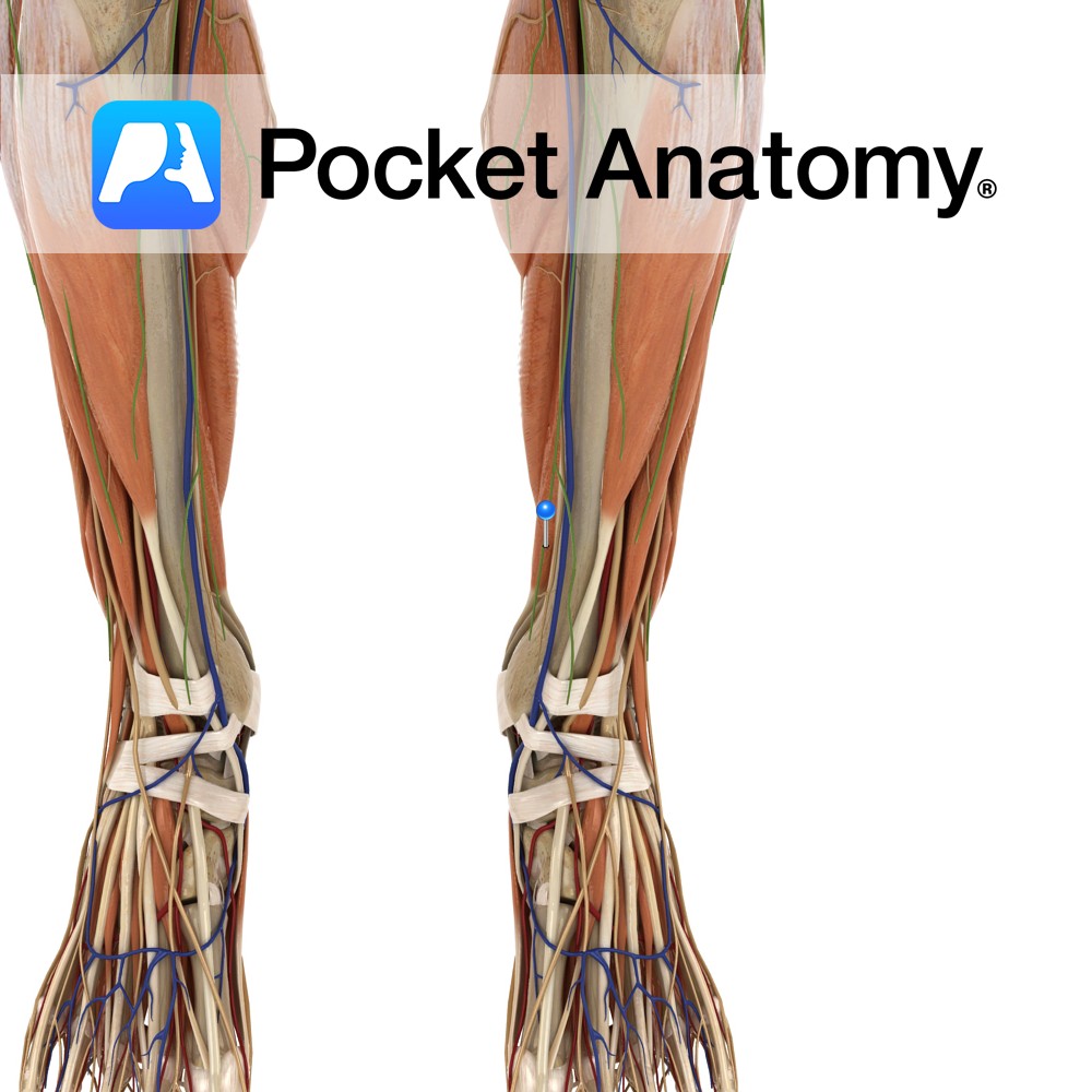
Medial side of the posterior surface of the tibia inferior to the soleal line.
Insertion:
Plantar surface of the base of the distal phalanges of the lateral four toes.
Key Relations:
-One of the four muscles of the deep posterior compartment of the leg.
-The flexor digitorum longus tendon passes posterior to the medial malleolus, deep to the flexor retinaculum and superficial to the flexor hallucis longus tendon.
Functions
-Flexes the lateral four toes e.g. gripping the ground with the toes.
-Weakly plantarflexes the ankle joint, with gastrocnemius.
-Active in maintaining the erect position during standing.
-Prevents excessive dorsiflexion during walking.
Supply
Nerve Supply:
Tibial nerve (L5, S1, S2).
Blood Supply:
–Posterior tibial artery
-Fibular (peroneal) artery.
Clinical
Flexor tendinitis is inflammation of the flexor tendons of the toes such as flexor digitorum longus and flexor hallucis longus. It most commonly affects dancers. Flexor tendinitis although often more painful is not as common as extensor tendinitis. The patient may present with pain and swelling along the course of the tendons, arch of the foot and inside of the ankle as well as increased pain with resisted flexion of the hallux (big toe).
Treatment includes rest, ice, NSAIDs and taping of the foot to support the arch. More severe cases may require surgery which has proven very successful with a recovery period of approximately 6 to 9 weeks.
Interested in taking our award-winning Pocket Anatomy app for a test drive?
![]()