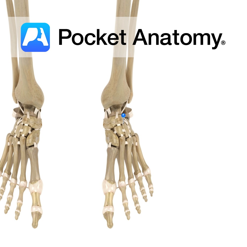
Motion
The various synovial joints between the individual tarsal bones invert, evert, supinate and pronate the foot. They allow the foot to adapt and maintain contact with the ground on irregular surfaces. The intertarsal joints are made up of the following individual articulations: Talocalcaneal, Talocalcanealnavicular, Calcaneocuboid, Cuneonavicular, Cubonavicular (normally fibrous), Intercuneiform, and Cuneocuboid.
The talocalcaneonavicular and calcaneocuboid together form what is commonly referered to as the ‘Transverse tarsal joint’. These 2 articulations along with the talocalcaneal articulation are responsible for most of the movements produced as the other joints have a very limited range of motion.
Stability
Talocalcaneal joint:
– The fibrous capsule of the joint contributes to its stability.
– Interosseus, lateral, medial, posterior and anterior ligamentsstabilize the joint.
Talocalcanealnavicular joint:
-The fibrous capsule of the joint contributes to its stability.
-Interosseus talocalcaneal (posteriorly), talonavicular (superiorly) and plantar calcaneonavicular (inferiorly) ligaments stabilize the joint. The talocalcaneonavicular ligament reinforces the joint laterally
Calcaneocuboid joint:
-The fibrous capsule of the joint contributes to its stability.
-Bifurcate, long plantar and plantar calcaneocuboid ligaments stabilize the joint.
Muscles
Inversion:
Tibialis anterior
Tibialis posterior
Eversion:
Peroneus longus
Peroneus Brevis.
Clinical
The bones involved in these joints may be affected by a number of pathologic processes including arthritis, gout, haemochromatosis.
Interested in taking our award-winning Pocket Anatomy app for a test drive?
![]()