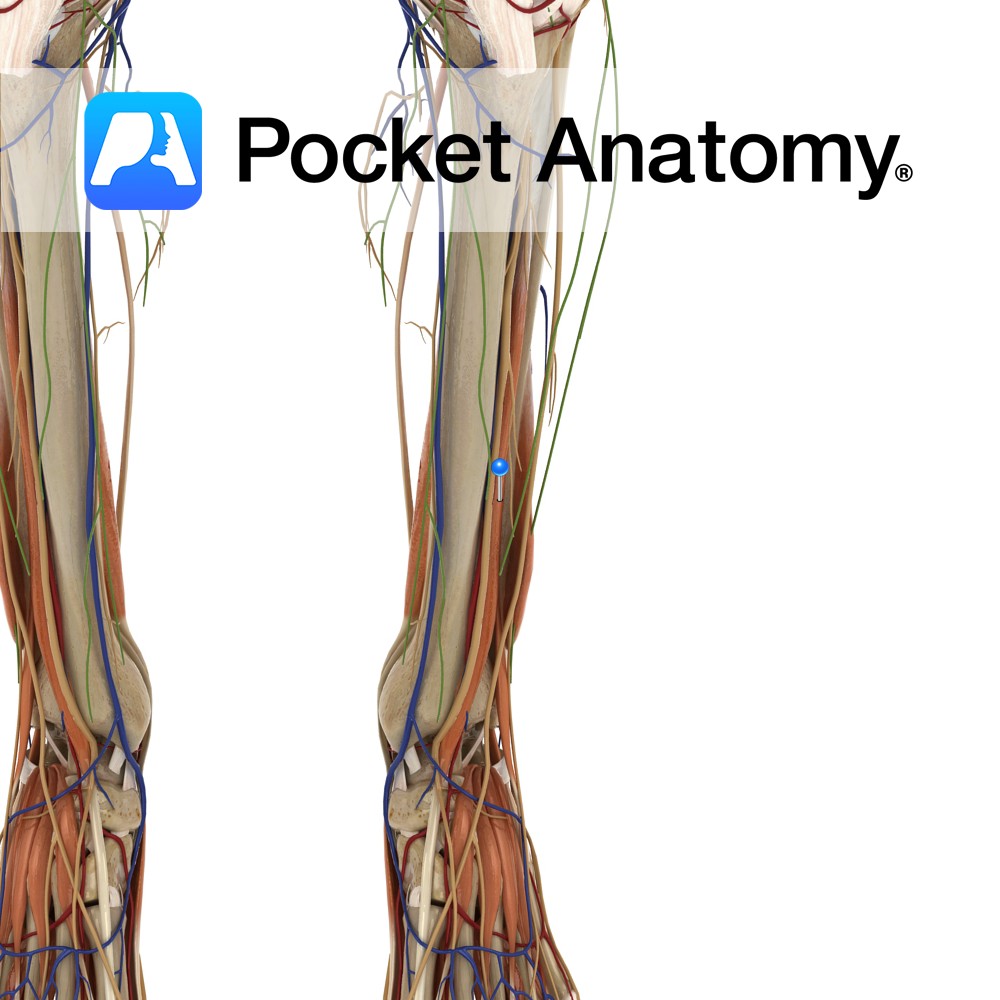
Anatomy
Origin:
Posterior surfaces of the tibia below the soleal line, posterior surface of the fibula and posterior surface of the interosseous membrane.
Insertion:
Tuberosity of the navicular, all cuneiforms, cuboid and the bases of the 2nd, 3rd and 4th metatarsals.
Key Relations:
-One of the four muscles of the deep posterior compartment of the leg.
-Travels infero-medially posterior to flexor digitorum longus. The tibialis posterior tendon passes posterior to the medial malleolus and inferior to the spring ligament.
Functions
-Inverts the foot.
-Plantarflexes the ankle joint e.g. pushing down the pedals when driving..
Supply
Nerve Supply:
Tibial nerve (L4, L5).
Blood Supply:
–Posterior tibial artery
-Fibular (peroneal) artery.
Clinical
Tibialis posterior syndrome is a rupture or overstretching of the muscle, leading to a fallen arch or flat foot, most commonly seen in middle-aged women. The patient may present with a fallen arch, possibly a history of injury to the muscle, with heel valgus, forefoot abduction and talus plantarflexion. Treatment includes strengthening of the tibialis posterior muscle, orthotics to support the arch and treatment of any associated conditions or injuries. More severe cases may require surgery to correct the deformity.
Interested in taking our award-winning Pocket Anatomy app for a test drive?
![]()