Anatomy
Course
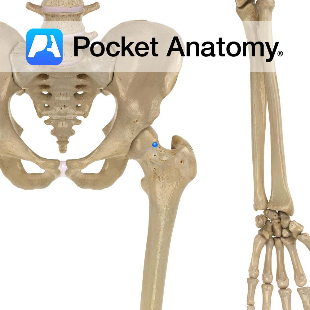
Originates from the deep femoral artery. It first passes between the iliopsoas and pectineus muscles, and then passes between the obturator externus and adductor brevis muscles.
Finally, it passes above the margin of the adductor magnus to travel medially around the neck of the femur. It then divides into two major branches deep to the quadratus femoris muscle. At the neck of the femur, it anastomoses with the other vessels at the head of the femur, to form part of the cruciate anastomosis.
Supply
Supplies the head and neck of the femur, as it anastomoses with the lateral circumflex artery.
Clinical
The medial circumflex artery and its branches are clinically relevant as it forms part of the cruciate anastomosis. Should a blockage arise between the internal iliac artery and the femoral artery, blood can reach the knee and lower leg via the cruciate anastomosis.
Interested in taking our award-winning Pocket Anatomy app for a test drive?
![]()