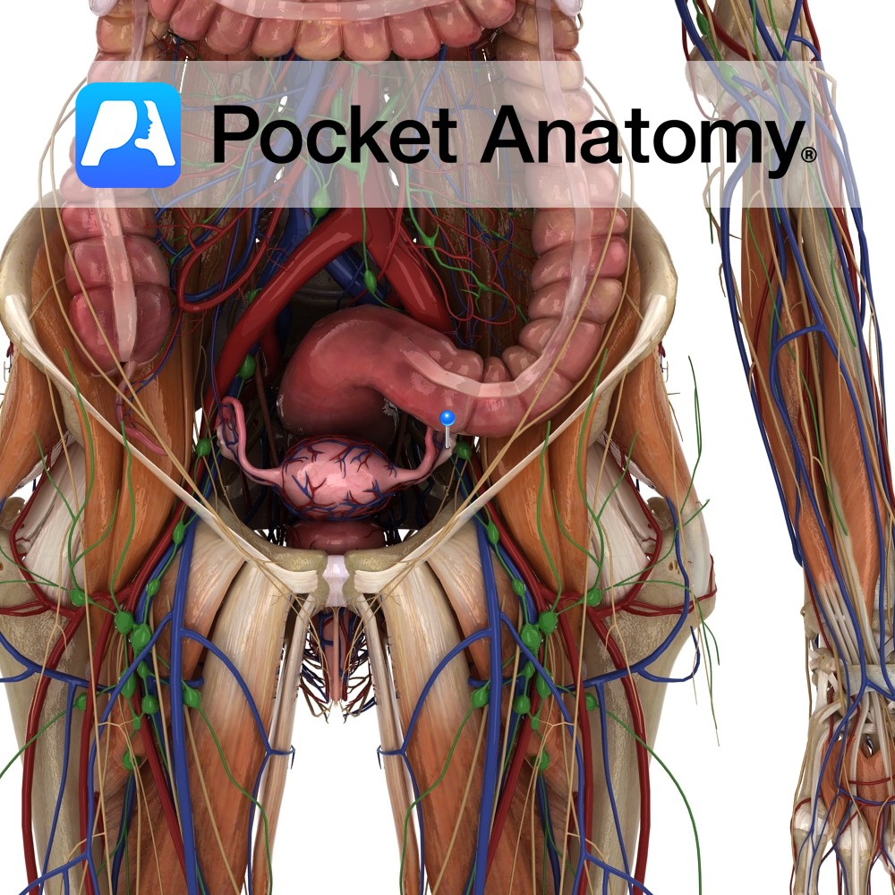
Anatomy
Female gonad (organ which secretes gametes – male/female secretes sperm/eggs). Paired, 4X2X1cm, 2-3.5gm (smaller with age from puberty), almond-shaped (long axis vertical), grey-pink, surface smooth (or puckered after repeated ovulation), sits at side of uterus, just below uterine/fallopian tube, in pelvic cavity, at or below level of umbilicus, in shallow ovarian fossa in lateral wall pelvis (in nulliparous/never-pregnant females; pushed/moved around by gravid uterus and subsequent location variable) beneath internal iliac artery, in front of ureter.
Held in place by ligaments (suspensory and ovarian) and by a fold of peritoneum (mesovarium) from the back of a greater fold (broad ligament) which envelopes the uterus and its various tubes. Upper/tubal extremity; covered by fimbriated infundibulum of fallopian tube, attached to suspensory ligament (through which course ovarian artery/vein, autonomic nerves, lymphatics to para-aortic nodes). Lower/uterine extremity connected to (lateral angle of) uterus by ovarian ligament (within broad ligament).
The tubal extremity: Upper end of ovary (ellipsoid shape with long axis vertical, though can change position, particularly after pregnancy, such that it lies horizontal rather than longitudinal), closely associated with greatly expanded lateral end of fallopian tube (fimbriated infundibulum), attached to pelvic wall by suspensory ligament of ovary (a peritoneal fold, being part of the upper border of the broad ligament, a doubled fold of peritoneum draped over and tucked around the uterus and associated structures) which carries the ovarian vessels.
The Uterine Extremity: Lower end of ovary (ellipsoid shape with long axis vertical, though can change position, particularly after pregnancy, such that it lies horizontal rather than longitudinal), attached to angle of uterus (ie where fallopian tube enters/joins uterus just below fundus) by cordlike ovarian ligament (as opposed to suspensory ligament of ovary) coursing between the 2 layers of the broad ligament.
Physiology
Females born with set number eggs (1-2,000,000, reducing to about 10% by age 30; boys continuously produce new sperm throughout life) in a stage of arrested meiosis (4N, prophase meiosis I) until puberty, when one egg (total of about 300-400 throughout life) wins the monthly race/competition (from menarche – onset of ovulation – to menopause – cessation) to development and release (ovulation, day 14 of a typical 28-day cycle), ordinarily finding its way into the nearer fallopian tube, where it may be fertilised by a male gamete (sperm) with the formation of a zygote, and travel on to implantation (about 5-9 days later) as a blastocyst (70-100 cells) in the uterus, constituting a pregnancy. A gland (makes and releases a substance) both exocrine (produces substance – ovum – and extrudes it into duct – fallopian tube) and endocrine (produces hormones – estrogen, progesterone – and secretes them directly/ductlessly into bloodstream). The only intra-peritoneal organ (ie not covered with peritoneum, as egg/ovum extruded/released from ovary – by rupture of a mature follicle which has made its way to some part of the ovarian surface – has to make its way through peritoneal “spacewalk” to enter the fallopian/uterine tube, which is not attached to the ovary).
Each month, from either the right or left ovary, an egg is released and ordinarily moves into the fallopian tube (encouraged by currents created by the wafting of fimbrial cilia), where it may be fertilised on meeting a sperm.
Interested in taking our award-winning Pocket Anatomy app for a test drive?
![]()

.jpg)