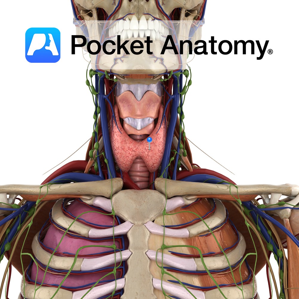
Anatomy
Largest endocrine gland, in neck, ductless, butterfly-shaped (2 conical lobes, apex up, connected by isthmus), behind infrahyoid (strap) and sternocleidomastoid muscles, partially wrapping around posterior structures – thyroid and cricoid cartilages (attached to them, so moves on swallowing), trachea, esophagus.
Encapsulated, between the 2 layers of which, posteriorly, are 2 parathyroid glands each side. Rich blood supply; superior (anastamosis right and left – external – common carotid – arch aorta) and inferior (thyrocervical – subclavian – arch aorta) thyroid arteries.
Thyroid veins; superior and middle – internal jugular, inferior (sometimes right and left join) – brachiocephalic. Internal structure; closed vesicles. Secretes hormones; T3/T4 (tri/tetra-iodothyronine, from tyrosine and iodine), calcitonin.
Interested in taking our award-winning Pocket Anatomy app for a test drive?
![]()
-%5Btrue-rib%5D.jpg)